Team:British Columbia/Model3
From 2011.igem.org
(Difference between revisions)
| Line 3: | Line 3: | ||
#bod {width:935px; float:left; background-color: white; margin-left: 15px; margin-top:10px;} | #bod {width:935px; float:left; background-color: white; margin-left: 15px; margin-top:10px;} | ||
</style><div id="bod"><center><h3>Monoterpene Synthase Structural Modeling</h3></center></html> | </style><div id="bod"><center><h3>Monoterpene Synthase Structural Modeling</h3></center></html> | ||
| - | <b>Objective:</b> To identify | + | <b>Objective:</b> To identify amino acid residues that are terpene synthesis, we constructed three-dimensional models of the terpene synthase proteins.<p></p> |
<b>Model Creators:</b> Joe Ho, Samuel Wu <p></p> | <b>Model Creators:</b> Joe Ho, Samuel Wu <p></p> | ||
| Line 15: | Line 15: | ||
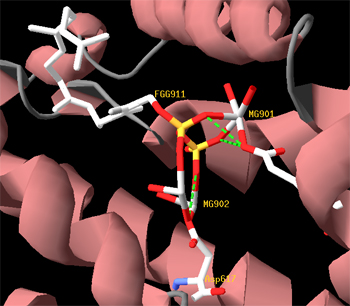
<b>Reactive site of the three synthases</b><p></p> | <b>Reactive site of the three synthases</b><p></p> | ||
[[File:UBCiGEM model3 reactive site.jpg]] | [[File:UBCiGEM model3 reactive site.jpg]] | ||
| + | Magnesium ions are cofactors located at the reactive site. | ||
<html><h3>Future Directions</h3></html><p></p> | <html><h3>Future Directions</h3></html><p></p> | ||
Revision as of 02:39, 29 October 2011

Monoterpene Synthase Structural Modeling
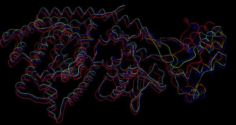
Three-dimensional structure of the 3 terpene synthases
Superimposition of the 3D structures of alpha-pinene synthase (blue), beta-pinene synthase (red), and limonene synthase (green). We used MODELLER (1) to automate homology-based 3D structure prediction. We identified an experimentally determined 3D structure of a taxadiene synthase from Pacific Yew (PDB ID: 3P5R) as an appropriate template. Reactive site of the three synthases Magnesium ions are cofactors located at the reactive site.
Future Directions
References
1. N. Eswar, M. A. Marti-Renom, B. Webb, M. S. Madhusudhan, D. Eramian, M. Shen, U. Pieper, A. Sali. Comparative Protein Structure Modeling With MODELLER. Current Protocols in Bioinformatics, John Wiley & Sons, Inc., Supplement 15, 5.6.1-5.6.30, 2006.
 "
"
