Monoterpene Synthase Structural Modeling
Objective: To identify amino acid residues that are terpene synthesis, we constructed three-dimensional models of the terpene synthase proteins.
Model Creators: Joe Ho, Samuel Wu
Model Advisors: Shing Hei Zhan, Alina Chan
Three-dimensional Structure of the 3 Terpene Synthase Proteins
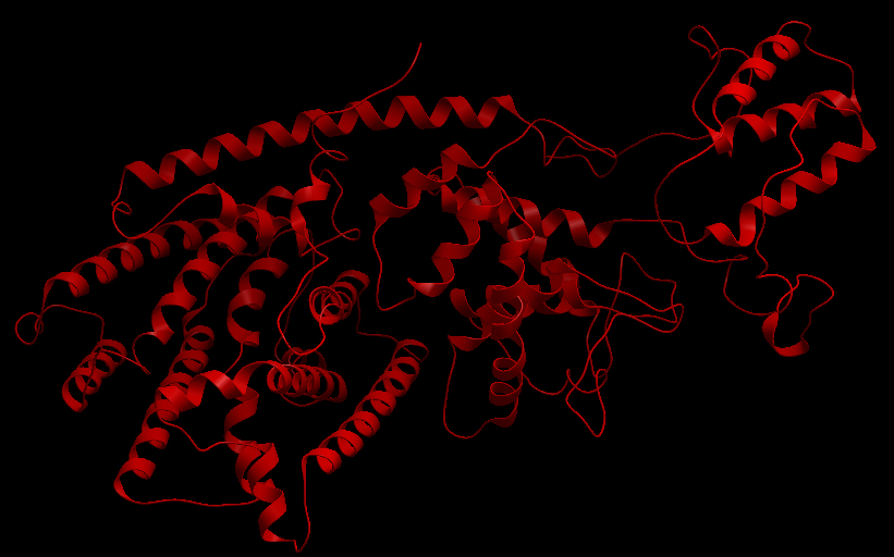
Ribbon representation of the alpha-pinene synthase.

Ribbon representation of the beta-pinene synthase.
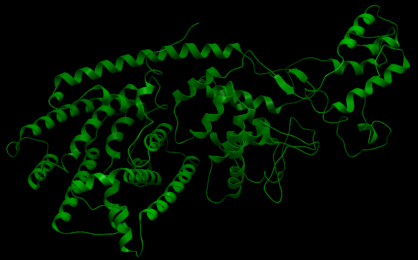
Ribbon representation of the limonene synthase.
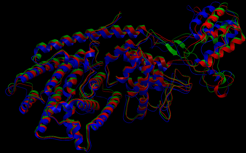
Superimposition of the 3D structures of the three synthase (blue), beta-pinene synthase (red), and limonene synthase (green). The synthases exhibit high structural similarity. This observation suggests that
Reactive site of the alpha-pinene synthase

Skin representation of the reactive site of alpha-pinene synthase. The substrate, geranyl diphosphate (GDP), is docked inside the reactive pocket. The oxygen atoms (red spheres) belonging to the phosphate groups of GDP and the carbon atoms (yellow spheres) forming the backbone of GDP are shown. Magnesium ion cofactors (blue spheres) interact with GDP.
Materials and Methods
The HQ426166, HQ426154, AY473624. We used MODELLER (1) to automate homology-based 3D structure prediction. We identified an experimentally determined 3D structure of a taxadiene synthase from Pacific Yew (PDB ID: 3P5R) as an appropriate template. The image was taken using the freely available ICM Browser.
Future Directions
References
1. N. Eswar, M. A. Marti-Renom, B. Webb, M. S. Madhusudhan, D. Eramian, M. Shen, U. Pieper, A. Sali. Comparative Protein Structure Modeling With MODELLER. Current Protocols in Bioinformatics, John Wiley & Sons, Inc., Supplement 15, 5.6.1-5.6.30, 2006.
 "
"


