 "
"
Team:Imperial College London/Reporters
From 2011.igem.org


Fluorescent Reporters

They can also be used for stunning visual effects.
Fluorescent reporters are an important tool in molecular biology, as they are frequently used to label various intracellular processes. In synthetic biology, fluorescent reporters are often used as the output of a genetic circuit, for example to signal the detection of a chemical.
As part of our iGEM project, we implemented a new fluorescent reporter, Dendra 2. In addition, we introduced a new coding sequence for superfolder GFP (sfGFP) that is codon-optimised for E. coli. These were both used as part of our imaging experiments of bacteria inside plant roots. sfGFP was also used in our soil experiments to label our bacteria so that we could later identify them.
sfGFP and mRFP were also used in our assembly strategy for the Gene Guard module. sfGFP was part of the construct of our CRIM plasmid, and would be used as a reporter to evaluate the level of expression of antiholin. mRFP formed part of the plasmid construct of the Gene Guard and this would be used to measure the expression of holin and endolysin. The mRFP would also be used to track the transmission of the plasmid in subsequent conjugation assays that would be carried out to test the effectiveness of the Gene Guard.

One of our cultures of E. coli expressing mRFP.
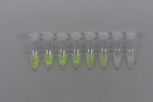
Samples of sfGFP from our thermostability assay. Note the colour change as the temperature increases from left to right.
Each of the fluorescent reporters that we studied was transformed into 5-alpha competent E. coli cells and then grown in large batch culture overnight. The cells were then lysed and the cell debris removed, leaving the protein suspended in 20 mM Tris buffer.
To further characterise these parts, we have conducted a thermostability assay to determine the temperature at which these proteins denature and cease to fluoresce. The data was collected in two overlapping sets, ranging from 35°C to 66°C, and from 57°C to 96°C. This was because it was only possible to fit a maximum of eight samples into the thermocycler at once.
Once the data was collected by taking fluorescence readings from a 96-well plate, it was normalised to a sample of untreated cell lysate containing the fluorescent protein. This gave a value for Relative Fluorescence, which was plotted against temperature to create a scatter plot. A line of best fit was applied, and the mid point of the sigmoid region of the line was taken as the denaturation point of the protein.
The protocols can be found in full on our Protocols page, which can be found by clicking on the button below.

Dendra 2
Dendra 2 is a green fluorescent protein that is capable of being irreversibly photo-converted by single-photon stimulation from excitation at 486 nm and emission at 505 nm wavelength to 558 nm excitation and 575nm emission wavelength. This means that its natural state is similar to GFP, but upon photo-conversion it can be excited as RFP. Photo-conversion can occur using two wavelengths 488 and 405 nm (Gurskaya et al., 2006)
Photo-conversion
Featured here are two of our videos demonstrating the photo-conversion of Dendra 2. Both videos show dendra expressing E.coli inside plant roots as they are photo-converted.
Video 1:A time-lapse video shows the conversion of cells in area 1. The single cell in area 3 serves as a negative control. It was not bleached by the laser and therefore continued to absorb light at a lower wavelength and emit green fluorescence.
How did we photo-convert Dendra 2 whilst exciting and detecting RFP?
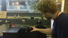
Tim uses his expertise to assist us.
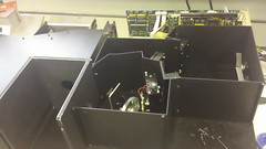
A look inside the fluorometer.
Using a FluoroMax-3 machine, and enlisting the expertise of Tim Wilson, we used Tim's 405 nm laser to photo-convert Dendra 2. This was done by aiming the laser into the sample cuvette from above.
So that we could excite Dendra 2 and measure its fluorescence as it was being photo-converted, Tim placed a filter taken from the Gel Imager in front of the detector so that we could detect the fluorescence from Dendra 2 without any interference from the excitation laser

This graph illustrates the photo-conversion of Dendra 2. The increase in fluorescence at 575 nm indicates that Dendra 2 begins to fluoresce red upon conversion by light at a wavelength of 405 nm. This also shows that the conversion is very rapid, which lends itself well to in vivo use as a fluorescent reporter.
Thermostability

This graph shows that Dendra 2 has a very high denaturation point at around 88°C. This lends the Dendra 2 protein very well to reporter assays where a high temperature is required. Our data also suggests that Dendra 2 may fluoresce more brightly as it is heated.
Superfolder GFP

This is a variant of GFP that has been engineered to be faster folding so that it can be used for tagging proteins more efficiently. The variant that we are submitting to the registry has been codon-optimised for E. coli.
Thermostability

From the midpoint of the sigmoidal curve, we can find the point at which half of the protein is denatured. For sfGFP, that point occurs at 78°C.
GFP

This GFP expression sequence is BioBrick BBa_I13522, from the Standard Registry of Biological Parts. This is a GFP coding sequence under the control of a pTet promoter. This was taken from the registry distribution and transformed into competent E. coli cells.
This helped to form the part of our project that required the re-categorisation of existing BioBricks as we carried out a thermostability assay.
Thermostability

mRFP

mRFP is a monomeric form of Red Fluorescent Protein, and the expression sequence that we used to obtain our samples is BioBrick BBa_I13521 from the Registry. This is a composite part made up of the coding sequence for mRFP under the control of a pTet promoter. This was taken from the registry distribution and transformed into competent E. coli cells.
We carried out a thermostability assay on mRFP in order to add the data to the registry data sheet.
Thermostability

CFP

CFP, or Cyan Fluorescent Protein, was taken from the registry as composite BioBrick BBa_I13600. This is the coding sequence for CFP under the control of a pTet promoter. This will give constitutive expression.