 "
"
Team:Imperial College London/Reporters
From 2011.igem.org


Fluorescent reporters
Dendra2
Dendra2 is the first photoconvertible protein that has been introduced into the Registry of Standard Biological Parts. When excited with a certain wavelength of light (405 nm or the less cytotoxic 488 nm), the protein is permanently converted from green to red. This interesting property allows it to be used for monitoring a variety of processes. In our experiments, we did preliminary experiments to test the use of Dendra2 to monitor the metabolic viability of our Escherichia coli once they are in the root.
Fig. 1: A time-lapse video shows the conversion of cells in area 1 (cluster of cells in the center of image). The single cell in area 3 (bottom right) serves as a negative control. It was not photoconverted by the laser and therefore continued to absorb light at a lower wavelength and emit green fluorescence. To find out more about this experiment, go to Phyto Route testing.
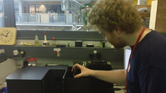
Fig. 2: Photo of Tim Wilson setting up the FluoroMax-3 machine for the Dendra2 photoconversion experiment.
Dendra2 has advantages over non-photoconvertible fluorescent proteins when used to monitor e.g. protein production and promoter characterisation. More accurate promoter characterisation will be possible since one would be able to photoconvert all of the Dendra2 and then measure the synthesis rate from point zero at different OD's.
From the literature [1] we found out that Dendra2 can be photoconverted using two wavelengths, however one of them (488 nm) is very close to excitation wavelength of green fluorescence (486 nm). Therefore we had to determine if the excitation wavelength (486 nm) for the Dendra2 before photoconversion would cause the Dendra2 to photoconvert. In order to test for that we excited cells with 486 nm & 558 nm wavelengths and measured the fluorescence at the emission wavelengths 505 nm and 575 nm.
Results for photostability of Dendra2:

Fig. 3:
We found that there was no RFP signal above that of background meaning that imaging the Dendra2 for GFP will not photoconvert the protein.
Once it was clear that the excitation wavelength does not interfere with the photoconversion wavelength we tested the photoconvertability of the Dendra2. In order to do that we used a FluoroMax-3 machine. Luckily, Tim Wilson was kind enough to help us set up the fluorometer in order to perform our experiments. We found that Dendra2 will fluoresce red upon conversion by light at a wavelength of 405 nm very quickly.
Results of photoconversion experiment:

Fig. 3: Results of the photoconversion experiment. Dendra2 is photoconverted with a wavelength of 405nm.
Thermostability assay
Fluorescent reporters are an important tool in synthetic biology that are often used as the output of a genetic circuit.
As part of our iGEM project, we implemented a new fluorescent reporter, Dendra2. In addition, we introduced a new coding sequence for superfolder GFP (sfGFP) that is codon-optimised for E. coli. These were both used as part of our imaging experiments of bacteria inside plant roots. sfGFP was also used in our soil experiments to label our bacteria so that we could later identify them.
sfGFP and mRFP were also used in our assembly strategy for the Gene Guard module. sfGFP was part of the construct of our CRIM plasmid, and would be used as a reporter to evaluate the level of expression of anti-holin. mRFP formed part of the plasmid construct of the Gene Guard and this would be used to measure the expression of holin and endolysin. The mRFP would also be used to track the transmission of the plasmid in subsequent conjugation assays that would be carried out to test the effectiveness of the Gene Guard.
Since we ended up working with so many fluorescent reporters during our project, we decided to further characterize these important BioBricks. An important characteristic that has been omitted from the registry page is the thermostability of these proteins. This is an important aspect when choosing a fluorescent protein for a thermophile.
To characterise these parts, we determined the temperature at which these proteins denature and cease to fluoresce. The data was collected in two overlapping sets, ranging from 35°C to 66°C, and from 57°C to 96°C. This was because it was only possible to fit a maximum of eight samples into the thermocycler at once.
Once the data was collected by taking fluorescence readings from a 96-well plate, it was normalised to a sample of untreated cell lysate containing the fluorescent protein. This gave a value for Relative Fluorescence, which was plotted against temperature to create a scatter plot. A line of best fit was applied, and the mid point of the sigmoid region of the line was taken as the denaturation point of the protein.

Fig. 4: Results of the heat denaturation experiment. The temperature at which half of the protein is denatured measured by looking at its fluorescence (PTm50) mRFP1: 92.3°C; GFPmut3b: 59.1°C; Dendra2: 83.7°C; sfGFP: 75.3°C.
References:
[1] Gurskaya, N.G. et al, (2006) Engineering of a monomeric green-to-red photoactivatable fluorescent protein induced by blue light. Nat Biotech, 24(4), pp.461-465.
<