Team:ETH Zurich/Overview/CircuitDesign
From 2011.igem.org
| Circuit Design |
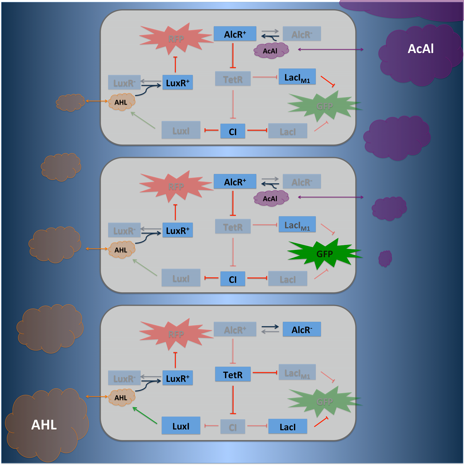
| To realize the smoke detecting optosensor (see Project Idea), we implemented two distinct network designs: one SmoColi sensing acethaldehyde (see Figure 1) and one sensing xylene (see Figure 2). Both designs share the same signal transduction and processing units, and a similar design in the signal detection unit. |
Signal Pre-ProcessingThe first step for both designs is to map an absolute acetaldehyde (xylene) concentration into a spatial signal. This first step is done in a channel by diffusion and degradation of acetaldehyde (xylene) a spatial gradient is produced. Signal Detection
Since for the further signal processing it was necessary to have a POPS signal proportional to the signaling molecule concentration, and since the acetaldehyde sensor inverts the signal due to its transcriptional inhibition, we decided to include an additional unit to invert the transduced signal downstream of acetaldehyde, which is not present for the xylene sensor: we put TetR under the transcriptional control of AlcR, and since TetR acts as a transcriptional inhibitor for downstream proteins, the transduced acetaldehyde signal has a similar qualitative input/output relation as the transduced xylene sensor without inverter. Signal Processing I: the Band-Pass FilterTo realize the spatial green fluorescence band indicating the acetaldehyde (xylene) concentration, we implemented a signal processing unit, which transduces the incoming signal if and only if the signal is neither too strong nor too weak (band-pass filter [2]): TetR represses (XylR enhances) the expression of the LacIM1 repressor (codon-modified LacI) and the lambda repressor CI. Thus, high acetaldehyde (xylene) concentrations result in high cytoplasmic levels of CI and of LacIM1. Since LacIM1 represses the expression of the green fluorescent protein (GFP), no green fluorescence signal can be detected for high concentrations. Low acetaldehyde (xylene) concentrations result in low LacIM1 and CI concentrations. Although in this case, LacIM1 does not repress GFP production, GFP is transcriptional repressed by the non-codon modified LacI, which is expressed at low CI concentrations. Only intermediate signals results in a moderate level of CI and LacIM1. The repression of CI is significantly higher than the one of LacIM1. While moderate levels of CI effectively shuts off LacI expression, the LacIM1 concentration is to low to repress GFP Signal Processing II: the Alarm SignalTo realize the alarm signal for toxic acetaldehyde (xylene) concentrations, we put the N-Acyl homoserine lactone (AHL) Synthase LuxI under the transcriptional control of CI, which is thus expressed only at low acetaldehyde (xylene) concentrations. LuxI syntesizes the small signaling molecule AHL which can quickly diffuse through the cell membrane in the extracellular space. Due to its relatively high diffusion constant, AHL diffuses through the whole tube and, thus, is present even in cells where no AHL is produced. Inside a cell, AHL can bind to its constitutively expressed receptor protein LuxR, and the thus activated LuxR represses the transcription of a red fluorescent protein (RFP). Thus, if the acetaldehyde concentration is below a threshold at least for a large enough minority of the cells in the channel, RFP is repressed in all cells. Only if the acetaldehyde concentration is above the threshold for all cells, no AHL is present in the channel and RFP is produced. Thus, the alarm signal RFP can be used to signal critical and potentially dangerous acetaldehyde (xylene) concentrations. |
 "
"


