Team:Bielefeld-Germany/Results/S-Layer/CspB CG
From 2011.igem.org
(→Identification and localization) |
(→Cultivation and protein expression) |
||
| (14 intermediate revisions not shown) | |||
| Line 14: | Line 14: | ||
===Cultivation and protein expression=== | ===Cultivation and protein expression=== | ||
| - | For characterization, PS2 also named CspB [http://partsregistry.org/Part:BBa_K525121 (K525121)] was fused with a monomeric RFP [http://partsregistry.org/Part:BBa_E1010 (BBa_E1010)] using [https://2011.igem.org/Team:Bielefeld-Germany/Protocols#Gibson_assembly Gibson assembly]. | + | For characterization, PS2, also named CspB [http://partsregistry.org/Part:BBa_K525121 (K525121)], was fused with a monomeric RFP [http://partsregistry.org/Part:BBa_E1010 (BBa_E1010)] using [https://2011.igem.org/Team:Bielefeld-Germany/Protocols#Gibson_assembly Gibson assembly]. |
The mRFP|CspB fusion protein was overexpressed in ''E. coli'' KRX after induction of T7 polymerase by supplementation of 0.1 % L-rhamnose using the [https://2011.igem.org/Team:Bielefeld-Germany/Protocols/Downstream-processing#Expression_of_S-layer_genes_in_E._coli autoinduction protocol]. | The mRFP|CspB fusion protein was overexpressed in ''E. coli'' KRX after induction of T7 polymerase by supplementation of 0.1 % L-rhamnose using the [https://2011.igem.org/Team:Bielefeld-Germany/Protocols/Downstream-processing#Expression_of_S-layer_genes_in_E._coli autoinduction protocol]. | ||
| - | [[Image:Bielefeld 2011 BF1 Growthcurve.png|450px|thumb|left| '''Figure 2: Growth curve of ''E. coli'' KRX expressing the fusion protein of CspB and mRFP with and without induction, cultivated at 37 °C in autoinduction medium with and without inductor, respectively. A curve depicting KRX wildtype is shown for | + | [[Image:Bielefeld 2011 BF1 Growthcurve.png|450px|thumb|left| '''Figure 2: Growth curve of ''E. coli'' KRX expressing the fusion protein of CspB and mRFP with and without induction, cultivated at 37 °C in autoinduction medium with and without inductor, respectively. A curve depicting KRX wildtype is shown for comparison. After autoinduction at approximately 4 h, a slight decrease in the OD<sub>600</sub> is observed approximately 4 hours later in the induced <partinfo>K525121</partinfo> compared to the uninduced culture. Both cultures grow significantly slower than KRX wildtype.]] |
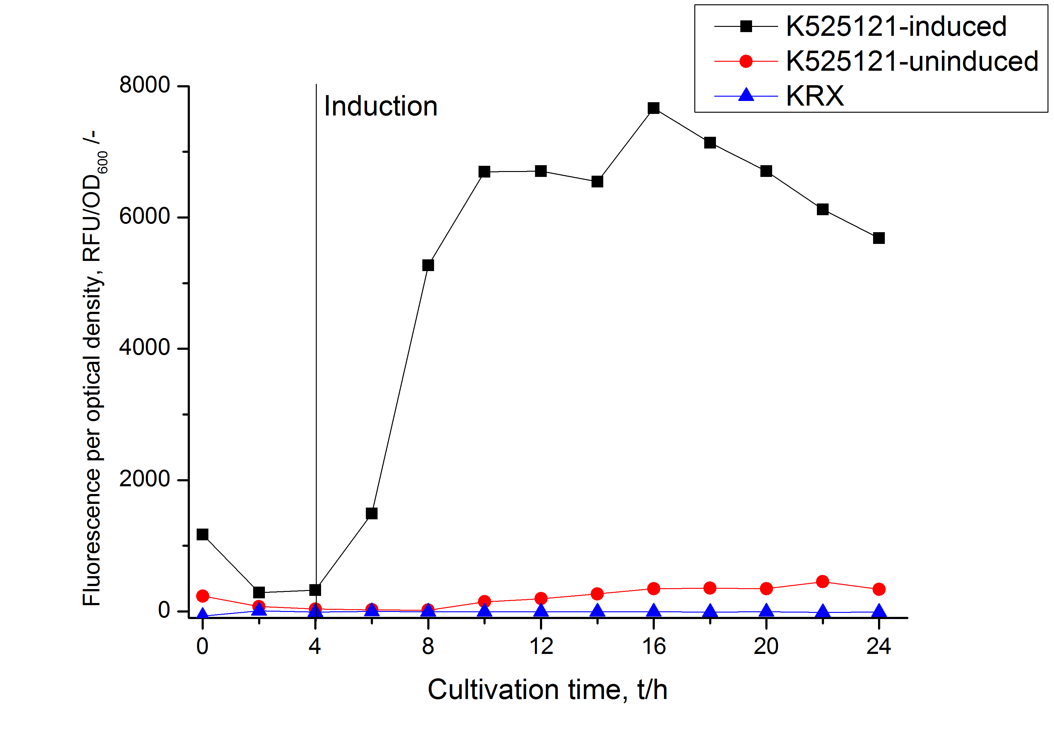
| - | [[Image:Bielefeld 2011 BF1 RFU OD.png|450px|thumb|right| '''Figure 3: RFU to OD<sub>600</sub> ratio of ''E. coli'' KRX expressing the fusion protein of CspB and mRFP with and without induction. A curve depicting KRX wildtype is shown for comparison. After induction at approximately 4 h the RFU to OD<sub>600</sub> ratio starts to rise in the induced culture. Compared to the uninduced culture the ratio is roughly seven times higher. The KRX wildtype shows no variation in the RFU to OD<sub>600</sub> ratio.]] | + | [[Image:Bielefeld 2011 BF1 RFU OD.png|450px|thumb|right| '''Figure 3: RFU to OD<sub>600</sub> ratio of ''E. coli'' KRX expressing the fusion protein of CspB and mRFP with and without induction. A curve depicting KRX wildtype is shown for comparison. After induction at approximately 4 h the RFU to OD<sub>600</sub> ratio starts to rise in the induced culture. Compared to the uninduced culture the ratio is roughly seven times higher. The KRX wildtype shows no variation in the RFU to OD<sub>600</sub> ratio.''']] |
<br style="clear: both" /> | <br style="clear: both" /> | ||
| Line 25: | Line 25: | ||
===Identification and localization=== | ===Identification and localization=== | ||
| - | [[Image:Bielefeld 2011 BF1 Purification.png|500px|thumb|right| '''Figure 4: Fluorescence progression of the mRFP [http://partsregistry.org/Part:BBa_E1010 (BBa_E1010)]/CspB fusion protein initiating with the cultivation fractions up to the detergent fractions of the | + | [[Image:Bielefeld 2011 BF1 Purification.png|500px|thumb|right| '''Figure 4: Fluorescence progression of the mRFP [http://partsregistry.org/Part:BBa_E1010 (BBa_E1010)]/CspB fusion protein initiating with the cultivation fractions up to the detergent fractions of the separate denaturations. Cultivations were carried out in autoinduction medium at 37 ˚C. The cells were mechanically disrupted and the resulting biomass was wahed with ddH<sub>2</sub>O and resuspendet in the respective detergent. The used detergent acronyms stand for: SDS = 10 % (v/v) sodium dodecyl sulfate; UTU = 7 M urea and 3 M thiourea; U = 10 M urea; NLS = 10 % (v/v) n-lauroyl sarcosine; CHAPS = 3-[(3-cholamidopropyl)dimethylammonio]-1-propanesulfonate.''']] |
| - | After a cultivation time of 18 h the mRFP|CspB fusion protein | + | After a cultivation time of 18 h the mRFP|CspB fusion protein the localization in ''E. coli'' KRX was analyzed. Therefore a part of the produced biomass was mechanically disrupted and the resulting lysate was washed with ddH<sub>2</sub>O. The periplasm was detached from other parts of the cells by using a osmotic shock. The existance of fluorescene in the periplasm fraction, shown in Fig. 4, indicates that ''C. glutamicum'' TAT-signal sequence is at least in part functional in ''E. coli'' KRX. |
The S-layer fusion protein could not be found in the lysate by SDS-PAGE and the cell debris were still red. This indicates that the fusion protein integrates into the cell membrane with it's lipid anchor. For testing this assumption the washed lysate was treated with ionic, nonionic and zwitterionic detergents to release the mRFP|CspB out of the membranes. | The S-layer fusion protein could not be found in the lysate by SDS-PAGE and the cell debris were still red. This indicates that the fusion protein integrates into the cell membrane with it's lipid anchor. For testing this assumption the washed lysate was treated with ionic, nonionic and zwitterionic detergents to release the mRFP|CspB out of the membranes. | ||
| - | The existance of fluorescence in the detergent fractions and the proportionally low fluorescence in the wash fraction compared to the lysis fraction confirm the hypothesis of an insertion into the cell membrane ( | + | The existance of fluorescence in the detergent fractions and the proportionally low fluorescence in the wash fraction compared to the lysis fraction confirm the hypothesis of an insertion into the cell membrane (Fig. 4). An insertion of these S-layer proteins might stabilize the membrane structure and increase the stability of cells against mechanical and chemical treatment. A stabilization of ''E. coli'' expressing S-layer proteins was described by [http://www.ncbi.nlm.nih.gov/pubmed/20829284 Lederer ''et al.'', (2010)]. |
| - | An other important fact is, that there is actually mRFP fluorescence measurable in such high concentrated detergent solutions. The S-layer seems to stabilize the biologically active conformation of mRFP. The MALDI-TOF analysis of the relevant size range in the polyacrylamid gel approved the existance of intact fusion protein in all detergent fractions ( | + | An other important fact is, that there is actually mRFP fluorescence measurable in such high concentrated detergent solutions. The S-layer seems to stabilize the biologically active conformation of mRFP. The MALDI-TOF analysis of the relevant size range in the polyacrylamid gel approved the existance of intact fusion protein in all detergent fractions (Fig. 5). |
<br style="clear: both" /> | <br style="clear: both" /> | ||
| - | MALDI-TOF analysis was first used to identify the location of the fusion protein in different fractions. Fractions of medium supernatant after cultivation, periplasmatic isolation, cell lysis, denaturation in 6 M urea and the following wash with 2 % (v/v) Triton X-100, 2 % SDS (w/v) were loaded onto a SDS-PAGE | + | MALDI-TOF analysis was first used to identify the location of the fusion protein in different fractions. Fractions of medium supernatant after cultivation, periplasmatic isolation, cell lysis, denaturation in 6 M urea and the following wash with 2 % (v/v) Triton X-100, 2 % SDS (w/v) were loaded onto a SDS-PAGE. The resulting gel was fragmented in slices containing proteins of similar size and analyzed with MALDI-TOF. |
[[Image:Bielefeld2011 K525131 page maldi.png|900px|thumb|center| '''Figure 5: SDS-PAGE of CspB/mRFP [http://partsregistry.org/Part:BBa_E1010 (BBa_E1010)] fusion protein. Lanes are fractions of culture supernatant (M), periplasmatic isolation (PP), cell lysis (L), denaturation (D) and a wash of the pellet of the denaturation with Triton X-100 (T). Used marker is PageRuler <sup>TM</sup> Prestained Protein Ladder SM0671. Marked regions were cut out and prepared for MALDI-TOF analysis.''']] | [[Image:Bielefeld2011 K525131 page maldi.png|900px|thumb|center| '''Figure 5: SDS-PAGE of CspB/mRFP [http://partsregistry.org/Part:BBa_E1010 (BBa_E1010)] fusion protein. Lanes are fractions of culture supernatant (M), periplasmatic isolation (PP), cell lysis (L), denaturation (D) and a wash of the pellet of the denaturation with Triton X-100 (T). Used marker is PageRuler <sup>TM</sup> Prestained Protein Ladder SM0671. Marked regions were cut out and prepared for MALDI-TOF analysis.''']] | ||
<br style="clear: both" /> | <br style="clear: both" /> | ||
| - | The following table shows the sequence coverage (in %) of our measurable gel samples with the amino acid sequence of fusion protein CspB | + | The following table shows the sequence coverage (in %) of our measurable gel samples with the amino acid sequence of fusion protein CspB|mRFP [http://partsregistry.org/Part:BBa_E1010 (BBa_E1010)]. |
<center> | <center> | ||
| Line 154: | Line 154: | ||
[[Image:Bielefeld2011_K525131_BF1_maldi_graph.png|500px|thumb|right| '''Figure 6: MALDI TOF measurement of CspB/mRFP [http://partsregistry.org/Part:BBa_E1010 (BBa_E1010)] fusion protein. Samples are arranged after estimated molecular mass of the gel slice. Measurement was performed with a ultrafleXtreme<sup>TM</sup> by Bruker Daltonics using the software FlexAnalysis, Biotools and SequenceEditor.''']] | [[Image:Bielefeld2011_K525131_BF1_maldi_graph.png|500px|thumb|right| '''Figure 6: MALDI TOF measurement of CspB/mRFP [http://partsregistry.org/Part:BBa_E1010 (BBa_E1010)] fusion protein. Samples are arranged after estimated molecular mass of the gel slice. Measurement was performed with a ultrafleXtreme<sup>TM</sup> by Bruker Daltonics using the software FlexAnalysis, Biotools and SequenceEditor.''']] | ||
| - | + | In Figure 6 sequence coverage data is visualized. The gel samples were arranged after estimated molecular mass cut out from the gel. As expected, only minor sequence coverage was found in the periplasmatic fraction, due to the lipid anchor located at the carboxy-terminus. This hydrophobic region inhibits the transport of the protein to the periplasm, mediated by the amino-terminal TAT-sequence. Little fluorescence was also found in the lysis fraction, verifying our assumtion, that the protein integrates or strongly binds to the cell membrane. Using urea to remove the S-layer fusion protein from the cell membrane resulted in a slightly higher sequence coverage. Washing the pellet with 2 % Triton X-100 (v/v), 2 % SDS (w/v), previously treated with urea, resulted in a higher sequence coverage and can therefore be expected as more applicable to desintegrate the S-layer fusion protein. The regarded sequence coverage in the supernatant of the cultivation medium may be caused by cell lysis during the late phase of cultivation. | |
<br style="clear: both" /> | <br style="clear: both" /> | ||
| - | To obtain more specific informations about the location of the S-layer fusion protein, after comparison with same treated fraction of ''E. coli'' KRX all gel bands in a defined size area were cut out of the gel and analysed with MALDI-TOF. Results are shown in | + | To obtain more specific informations about the location of the S-layer fusion protein, after comparison with same treated fraction of ''E. coli'' KRX all gel bands in a defined size area were cut out of the gel and analysed with MALDI-TOF. Results are shown in Fig. 7. |
| - | [[Image:Bielefeld2011_K525131_BF1_Gel1.png|900px|thumb|center| '''Figure 7: MALDI-TOF measurement of CspB/mRFP [http://partsregistry.org/Part:BBa_E1010 (BBa_E1010)] fusion protein in different fractions. Abbreviations are Ma: Marker (PageRuler <sup>TM</sup> Prestained Protein Ladder SM0671), M (medium), PP (periplasm), L (cell lysis with ribolyser), W (wash with ddH<sub>2</sub>O). In the left half of the gel fractions of ''E. coli'' KRX with induced production of fusion protein, the right half shows fractions of ''E. coli'' KRX without carrying the plasmid coding the fusion protein. Colours show the sequence coverage of the gel lane, | + | [[Image:Bielefeld2011_K525131_BF1_Gel1.png|900px|thumb|center| '''Figure 7: MALDI-TOF measurement of CspB/mRFP [http://partsregistry.org/Part:BBa_E1010 (BBa_E1010)] fusion protein in different fractions. Abbreviations are Ma: Marker (PageRuler <sup>TM</sup> Prestained Protein Ladder SM0671), M (medium), PP (periplasm), L (cell lysis with ribolyser), W (wash with ddH<sub>2</sub>O). In the left half of the gel fractions of ''E. coli'' KRX with induced production of fusion protein, the right half shows fractions of ''E. coli'' KRX without carrying the plasmid coding the fusion protein. Colours show the sequence coverage of the gel lane, cut out of the gel.''']] |
<br style="clear: both" /> | <br style="clear: both" /> | ||
| - | Sequence coverage was only found in the wash and the lysis fraction, | + | Sequence coverage was only found in the wash and the lysis fraction, assures the assumption that the S-layer protein is integrating in the cell membrane. |
The influence of other detergents to disintegrate the S-layer fusion protein was tested after disrupting the cells with a ribolyser. The cell pellet was incubated in 10 % (v/v) Sodium dodecyl sulfate (SDS), in 7 M urea and 3 M thiourea (UTU), in 10 M urea (U) in 10 % (v/v) n-lauroyl sarcosine (NLS) and in 2 % CHAPS (C). Samples of the incubations with these detergents were loaded onto a SDS-PAGE prior to measurement with MALDI TOF. | The influence of other detergents to disintegrate the S-layer fusion protein was tested after disrupting the cells with a ribolyser. The cell pellet was incubated in 10 % (v/v) Sodium dodecyl sulfate (SDS), in 7 M urea and 3 M thiourea (UTU), in 10 M urea (U) in 10 % (v/v) n-lauroyl sarcosine (NLS) and in 2 % CHAPS (C). Samples of the incubations with these detergents were loaded onto a SDS-PAGE prior to measurement with MALDI TOF. | ||
| Line 178: | Line 178: | ||
===Cultivation and protein expression=== | ===Cultivation and protein expression=== | ||
| - | For characterization the modiefied CspB [http://partsregistry.org/Part:BBa_K525123 (K525123)] gen was fused with a monomeric RFP [http://partsregistry.org/Part:BBa_E1010 (BBa_E1010)] using [https://2011.igem.org/Team:Bielefeld-Germany/Protocols#Gibson_assembly Gibson assembly]. | + | For characterization, the modiefied CspB [http://partsregistry.org/Part:BBa_K525123 (K525123)] gen was fused with a monomeric RFP [http://partsregistry.org/Part:BBa_E1010 (BBa_E1010)] using [https://2011.igem.org/Team:Bielefeld-Germany/Protocols#Gibson_assembly Gibson assembly]. |
The fusion protein was overexpressed in ''E. coli'' KRX after induction of T7 polymerase by supplementation of 0,1 % L-rhamnose using the [https://2011.igem.org/Team:Bielefeld-Germany/Protocols/Downstream-processing#Expression_of_S-layer_genes_in_E._coli autoinduction protocol]. | The fusion protein was overexpressed in ''E. coli'' KRX after induction of T7 polymerase by supplementation of 0,1 % L-rhamnose using the [https://2011.igem.org/Team:Bielefeld-Germany/Protocols/Downstream-processing#Expression_of_S-layer_genes_in_E._coli autoinduction protocol]. | ||
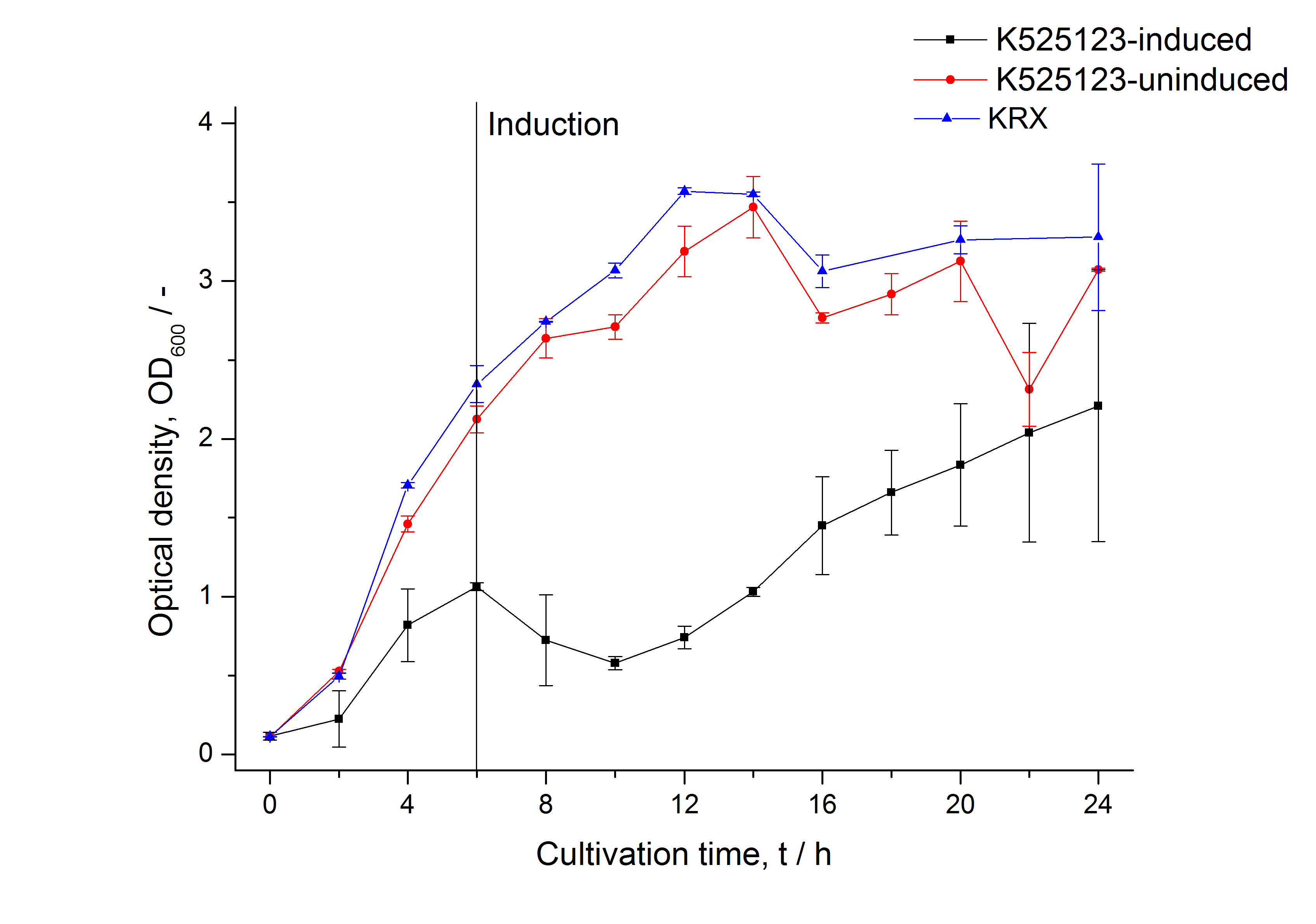
| - | [[Image: Bielefeld 2011 BF3 Growthcurve.png|450px|thumb|left| '''Figure 9: Growthcurve of ''E. coli'' KRX expressing the fusion protein of CspB and mRFP with and without induction, cultivated at 37 °C in autoinduction medium with, respectively, without inductor. A curve depicting KRX wildtype is shown for | + | [[Image: Bielefeld 2011 BF3 Growthcurve.png|450px|thumb|left| '''Figure 9: Growthcurve of ''E. coli'' KRX expressing the fusion protein of CspB and mRFP with and without induction, cultivated at 37 °C in autoinduction medium with, respectively, without inductor. A curve depicting KRX wildtype is shown for comparison. After induction at approximately 6 h the OD<sub>600</sub> of the induced K525123 visibly drops when compared to the uninduced culture. While the induced culture grow significantly slower than KRX wildtype the uninduced seems to be unaffected.]] |
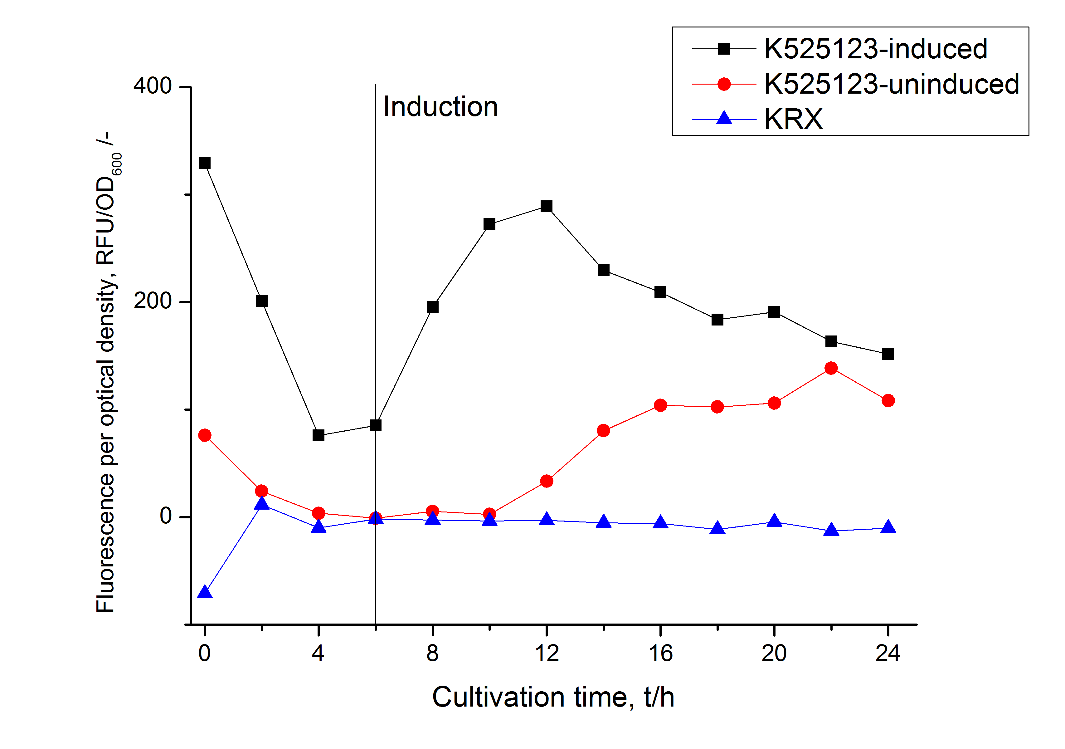
| - | [[Image: Bielefeld 2011 BF3 RFU OD.png|450px|thumb|right| '''Figure 10: RFU to OD<sub>600</sub> ratio of ''E. coli'' KRX expressing the fusion protein of CspB and mRFP with and without induction. A curve depicting KRX wildtype is shown for | + | [[Image: Bielefeld 2011 BF3 RFU OD.png|450px|thumb|right| '''Figure 10: RFU to OD<sub>600</sub> ratio of ''E. coli'' KRX expressing the fusion protein of CspB and mRFP with and without induction. A curve depicting KRX wildtype is shown for comparison. After induction at approximately 6 h the RFU to OD<sub>600</sub> ratio starts to rise in the induced culture. Compared to the uninduced culture the ratio is roughly eight times higher. Most likely due to basal transcription the RFU to OD<sub>600</sub> ratio of the uninduced culture starts to rise after 12 hours. The KRX wildtype shows no variation in the RFU to OD<sub>600</sub> ratio.]] |
<br style="clear: both" /> | <br style="clear: both" /> | ||
| - | ===Identification and | + | ===Identification and localization=== |
| - | [[Image:Bielefeld 2011 BF3 Purification.png|500px|thumb|right| '''Figure 11: Fluorescence progression of the mRFP[http://partsregistry.org/Part:BBa_E1010 (BBa_E1010)]/CspB fusion protein initiating with the cultivation fractions up to the detergent fractions of the | + | [[Image:Bielefeld 2011 BF3 Purification.png|500px|thumb|right| '''Figure 11: Fluorescence progression of the mRFP[http://partsregistry.org/Part:BBa_E1010 (BBa_E1010)]/CspB fusion protein initiating with the cultivation fractions up to the detergent fractions of the separate denaturations. Cultivations were carried out in autoinduction medium at 37 ˚C. The cells were mechanically disrupted and the resulting biomass was washed with ddH<sub>2</sub>O and resuspended in the respective detergent. The used detergent acronyms stand for: SDS = 10 % sodium dodecyl sulfate; UTU = 7 M urea and 3 M thiourea; U = 10 M urea; NLS = 10 % n-lauroyl sarcosine; 2 % CHAPS = 3-[(3-cholamidopropyl)dimethylammonio]-1-propanesulfonate.''']] |
| - | After a cultivation time of 18 h the mRFP|CspB fusion protein | + | After a cultivation time of 18 h the mRFP|CspB fusion protein the localization in ''E. coli'' KRX was analyzed. Therefor a part of the produced biomass was mechanically disrupted and the resulting lysate was washed with ddH<sub>2</sub>O. The periplasm was detached by using an osmotic shock. |
The S-layer fusion protein could not be found in the polyacrylamide gel after a SDS-PAGE of the lysate. This indicated that the fusion protein integrates into the cell membrane with its lipid anchor. For testing this assumption the washed lysate was treated with ionic, nonionic and zwitterionic detergents to release the mRFP|CspB out of the membranes. | The S-layer fusion protein could not be found in the polyacrylamide gel after a SDS-PAGE of the lysate. This indicated that the fusion protein integrates into the cell membrane with its lipid anchor. For testing this assumption the washed lysate was treated with ionic, nonionic and zwitterionic detergents to release the mRFP|CspB out of the membranes. | ||
| - | The existance of | + | The existance of fluorescence in the detergent fractions and the not existent fluorescence in the wash fraction confirms the hypothesis of an insertion into the cell membrane (Fig. 11). An insertion of these S-layer proteins might stabilize the membrane structure and increase the stability of cells against mechanical and chemical treatment. A stabilization of ''E. coli'' expressing S-layer proteins was described by [http://www.ncbi.nlm.nih.gov/pubmed/20829284 Lederer ''et al.'', (2010)]. |
| - | Another important fact is that there is actually mRFP fluorescence measurable in such high concentrated detergent solutions. The S-layer seems to stabilize the biologically active conformation of mRFP. The MALDI-TOF analysis of the relevant size range in the polyacrylamid gel approved the existance of the intact fusion protein in all detergent fractions ( | + | Another important fact is that there is actually mRFP fluorescence measurable in such high concentrated detergent solutions. The S-layer seems to stabilize the biologically active conformation of mRFP. The MALDI-TOF analysis of the relevant size range in the polyacrylamid gel approved the existance of the intact fusion protein in all detergent fractions (Fig. 12). |
| - | In comparison with the mRFP fusion protein of [http://partsregistry.org/Part:BBa_K525121 K525121], which has a TAT-sequence, a minor relative fluorescence in all cultivation and detergent fractions was detected ( | + | In comparison with the mRFP fusion protein of [http://partsregistry.org/Part:BBa_K525121 K525121], which has a TAT-sequence, a minor relative fluorescence in all cultivation and detergent fractions was detected (Fig. 11). Together with the decreasing RFU/OD<sub>600</sub> after 12 h of cultivation (Fig. 10) indicates that the TAT-sequence results in a postive effect on the protein stability. |
<br style="clear: both" /> | <br style="clear: both" /> | ||
| - | MALDI-TOF analysis was used to identify the location of the fusion protein in different fractions. Fractions of medium supernatant after cultivation (M), periplasmatic isolation (PP), cell lysis (L) and the following wash with ddH<sub>2</sub>O, samples were loaded onto a SDS-PAGE. After comparison with same treated fraction of E. coli KRX all gel bands in a defined size area were | + | MALDI-TOF analysis was used to identify the location of the fusion protein in different fractions. Fractions of medium supernatant after cultivation (M), periplasmatic isolation (PP), cell lysis (L) and the following wash with ddH<sub>2</sub>O, samples were loaded onto a SDS-PAGE. After comparison with same treated fraction of E. coli KRX all gel bands in a defined size area were cut out of the gel and analysed with MALDI-TOF. Results are shown in Fig. 12. |
| Line 213: | Line 213: | ||
Results show that the fusion protein of mRFP[http://partsregistry.org/Part:BBa_E1010 (BBa_E1010)]/CspB without TAT-sequence and with lipid anchor has only been identified in the lysis fraction. However, in conclusion with absent TAT-sequence, the protein has not been identified in the periplasm and the culture supernatant, respectively. | Results show that the fusion protein of mRFP[http://partsregistry.org/Part:BBa_E1010 (BBa_E1010)]/CspB without TAT-sequence and with lipid anchor has only been identified in the lysis fraction. However, in conclusion with absent TAT-sequence, the protein has not been identified in the periplasm and the culture supernatant, respectively. | ||
| - | The influence of other detergents to disintegrate the S-layer fusion protein was tested after disrupting the cells with a ribolyser. The cell pellet was incubated in 10 % (v/v) Sodium dodecyl sulfate (SDS), in 7 M urea and 3 M thiourea (UTU), in 10 M urea (U) in 10 % (v/v) N-lauroyl sarcosine (NLS) and in 2 % CHAPS (C). Samples of the incubations with these detergents were loaded onto a SDS-PAGE prior to measurement with MALDI-TOF ( | + | The influence of other detergents to disintegrate the S-layer fusion protein was tested after disrupting the cells with a ribolyser. The cell pellet was incubated in 10 % (v/v) Sodium dodecyl sulfate (SDS), in 7 M urea and 3 M thiourea (UTU), in 10 M urea (U) in 10 % (v/v) N-lauroyl sarcosine (NLS) and in 2 % CHAPS (C). Samples of the incubations with these detergents were loaded onto a SDS-PAGE prior to measurement with MALDI-TOF (Fig. 13). |
Latest revision as of 03:46, 29 October 2011


Contents |
CspB from Corynebacterium glutamicum
The S-layer of the gram-positive bacterium Corynebacterium glutamicum ATCC 14067 is formed by the PS2 (alternatively CspB) protein. The protein is encoded by the gene cspB. The mature protein has a molecular mass of 52.5 kDa. It is devoid of any sulfur-containing amino acids, whereas its nature is due to a high content of hydrophobic amino acids. Although a lot of different S-layer proteins exist, PS2 has no similarities to any other protein in the EMBL database. The S-layer of C. glutamicum is characterized by a hexagonal lattice symmetry (see Fig. 1). Attachment between S-layer and cell wall was found to be due to the hydrophobic carboxy-terminus of the PS2 protein. It was found that peptidoglycan is probably not involved in the interaction between the PS2 S-layer and the cell because the interaction between PS2 and the cell is disrupted by adding detergents. Also the S-layer protein from C. glutamicum does not contain a SLH domain, which is characteristic for several S-layer proteins and other enzymes bound to peptidoglycan. Besides, some other S-layer proteins show a carboxy-terminal hydrophobic sequence of 20 – 24 amino acids. (e.g. Halobacterium halobium, Haloferax volcanii, Rickettsia rickettsii) ([http://onlinelibrary.wiley.com/doi/10.1046/j.1365-2958.1997.d01-1868.x/abstract Chami et al., 1997], [http://www.sciencedirect.com/science/article/pii/S016816560400241X Hansmeier et al., 2004]).
CspB with TAT-sequence and lipid anchor
Cultivation and protein expression
For characterization, PS2, also named CspB [http://partsregistry.org/Part:BBa_K525121 (K525121)], was fused with a monomeric RFP [http://partsregistry.org/Part:BBa_E1010 (BBa_E1010)] using Gibson assembly.
The mRFP|CspB fusion protein was overexpressed in E. coli KRX after induction of T7 polymerase by supplementation of 0.1 % L-rhamnose using the autoinduction protocol.

Identification and localization
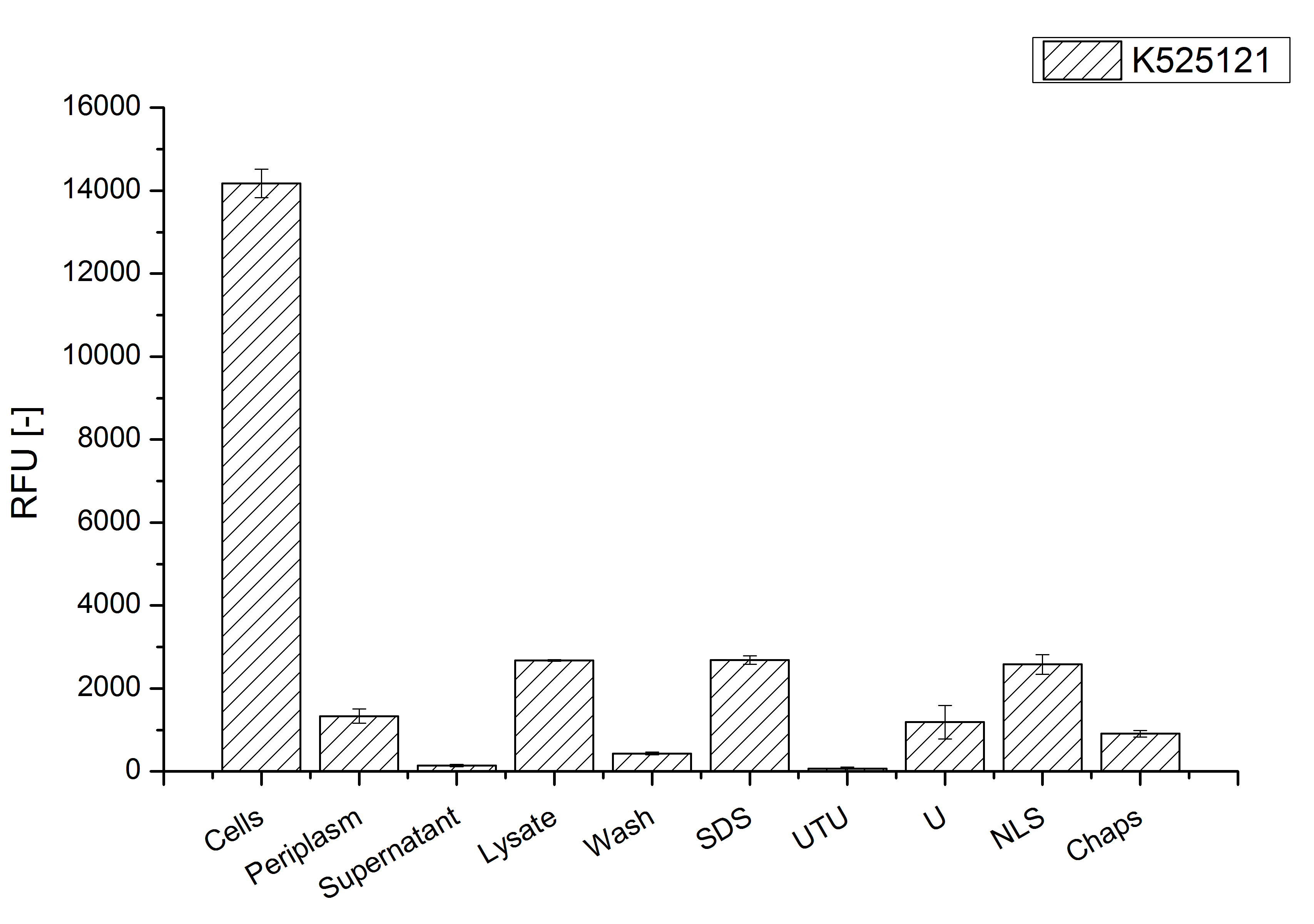
After a cultivation time of 18 h the mRFP|CspB fusion protein the localization in E. coli KRX was analyzed. Therefore a part of the produced biomass was mechanically disrupted and the resulting lysate was washed with ddH2O. The periplasm was detached from other parts of the cells by using a osmotic shock. The existance of fluorescene in the periplasm fraction, shown in Fig. 4, indicates that C. glutamicum TAT-signal sequence is at least in part functional in E. coli KRX.
The S-layer fusion protein could not be found in the lysate by SDS-PAGE and the cell debris were still red. This indicates that the fusion protein integrates into the cell membrane with it's lipid anchor. For testing this assumption the washed lysate was treated with ionic, nonionic and zwitterionic detergents to release the mRFP|CspB out of the membranes.
The existance of fluorescence in the detergent fractions and the proportionally low fluorescence in the wash fraction compared to the lysis fraction confirm the hypothesis of an insertion into the cell membrane (Fig. 4). An insertion of these S-layer proteins might stabilize the membrane structure and increase the stability of cells against mechanical and chemical treatment. A stabilization of E. coli expressing S-layer proteins was described by [http://www.ncbi.nlm.nih.gov/pubmed/20829284 Lederer et al., (2010)].
An other important fact is, that there is actually mRFP fluorescence measurable in such high concentrated detergent solutions. The S-layer seems to stabilize the biologically active conformation of mRFP. The MALDI-TOF analysis of the relevant size range in the polyacrylamid gel approved the existance of intact fusion protein in all detergent fractions (Fig. 5).
MALDI-TOF analysis was first used to identify the location of the fusion protein in different fractions. Fractions of medium supernatant after cultivation, periplasmatic isolation, cell lysis, denaturation in 6 M urea and the following wash with 2 % (v/v) Triton X-100, 2 % SDS (w/v) were loaded onto a SDS-PAGE. The resulting gel was fragmented in slices containing proteins of similar size and analyzed with MALDI-TOF.

The following table shows the sequence coverage (in %) of our measurable gel samples with the amino acid sequence of fusion protein CspB|mRFP [http://partsregistry.org/Part:BBa_E1010 (BBa_E1010)].
| number of gel sample | sequence coverage (%) | |
|---|---|---|
| 1 | 1.9 | |
| 2 | 11.5 | |
| 3 | 8.0 | |
| 4 | 2.6 | |
| 5 | 0.0 | |
| 6 | 0.0 | |
| 7 | 2.6 | |
| 8 | 0.0 | |
| 9 | 0.0 | |
| 10 | 0.0 | |
| 11 | 9.4 | |
| 12 | 2.6 | |
| 13 | 2.8 | |
| 14 | 0.0 | |
| 15 | 0.0 | |
| 16 | 0.0 | |
| 17 | 8.0 | |
| 18 | 0.0 | |
| 19 | 0.0 | |
| 20 | 0.0 | |
| 21 | 12.2 | |
| 22 | 12.2 | |
| 23 | 0.0 | |
| 24 | 0.0 | |
| 25 | 0.0 |
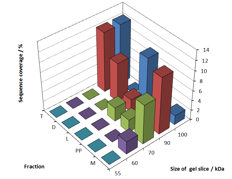
In Figure 6 sequence coverage data is visualized. The gel samples were arranged after estimated molecular mass cut out from the gel. As expected, only minor sequence coverage was found in the periplasmatic fraction, due to the lipid anchor located at the carboxy-terminus. This hydrophobic region inhibits the transport of the protein to the periplasm, mediated by the amino-terminal TAT-sequence. Little fluorescence was also found in the lysis fraction, verifying our assumtion, that the protein integrates or strongly binds to the cell membrane. Using urea to remove the S-layer fusion protein from the cell membrane resulted in a slightly higher sequence coverage. Washing the pellet with 2 % Triton X-100 (v/v), 2 % SDS (w/v), previously treated with urea, resulted in a higher sequence coverage and can therefore be expected as more applicable to desintegrate the S-layer fusion protein. The regarded sequence coverage in the supernatant of the cultivation medium may be caused by cell lysis during the late phase of cultivation.
To obtain more specific informations about the location of the S-layer fusion protein, after comparison with same treated fraction of E. coli KRX all gel bands in a defined size area were cut out of the gel and analysed with MALDI-TOF. Results are shown in Fig. 7.

Sequence coverage was only found in the wash and the lysis fraction, assures the assumption that the S-layer protein is integrating in the cell membrane.
The influence of other detergents to disintegrate the S-layer fusion protein was tested after disrupting the cells with a ribolyser. The cell pellet was incubated in 10 % (v/v) Sodium dodecyl sulfate (SDS), in 7 M urea and 3 M thiourea (UTU), in 10 M urea (U) in 10 % (v/v) n-lauroyl sarcosine (NLS) and in 2 % CHAPS (C). Samples of the incubations with these detergents were loaded onto a SDS-PAGE prior to measurement with MALDI TOF.

The result of the MALDI-TOF measurement clearly demonstrates that all used detergents are applicable to disintegrate the S-layer fusion proteins from the bacterial cell membrane of E. coli. Fluorescence measurement of fractions, treated with the detergents, show significantly different values, indicating that some of the detergents (e.g. 3 M thiourea, 7 M urea) have a strong effect on protein folding.
CspB without TAT-sequence and with lipid anchor
Cultivation and protein expression
For characterization, the modiefied CspB [http://partsregistry.org/Part:BBa_K525123 (K525123)] gen was fused with a monomeric RFP [http://partsregistry.org/Part:BBa_E1010 (BBa_E1010)] using Gibson assembly.
The fusion protein was overexpressed in E. coli KRX after induction of T7 polymerase by supplementation of 0,1 % L-rhamnose using the autoinduction protocol.
Identification and localization

After a cultivation time of 18 h the mRFP|CspB fusion protein the localization in E. coli KRX was analyzed. Therefor a part of the produced biomass was mechanically disrupted and the resulting lysate was washed with ddH2O. The periplasm was detached by using an osmotic shock.
The S-layer fusion protein could not be found in the polyacrylamide gel after a SDS-PAGE of the lysate. This indicated that the fusion protein integrates into the cell membrane with its lipid anchor. For testing this assumption the washed lysate was treated with ionic, nonionic and zwitterionic detergents to release the mRFP|CspB out of the membranes.
The existance of fluorescence in the detergent fractions and the not existent fluorescence in the wash fraction confirms the hypothesis of an insertion into the cell membrane (Fig. 11). An insertion of these S-layer proteins might stabilize the membrane structure and increase the stability of cells against mechanical and chemical treatment. A stabilization of E. coli expressing S-layer proteins was described by [http://www.ncbi.nlm.nih.gov/pubmed/20829284 Lederer et al., (2010)].
Another important fact is that there is actually mRFP fluorescence measurable in such high concentrated detergent solutions. The S-layer seems to stabilize the biologically active conformation of mRFP. The MALDI-TOF analysis of the relevant size range in the polyacrylamid gel approved the existance of the intact fusion protein in all detergent fractions (Fig. 12).
In comparison with the mRFP fusion protein of [http://partsregistry.org/Part:BBa_K525121 K525121], which has a TAT-sequence, a minor relative fluorescence in all cultivation and detergent fractions was detected (Fig. 11). Together with the decreasing RFU/OD600 after 12 h of cultivation (Fig. 10) indicates that the TAT-sequence results in a postive effect on the protein stability.
MALDI-TOF analysis was used to identify the location of the fusion protein in different fractions. Fractions of medium supernatant after cultivation (M), periplasmatic isolation (PP), cell lysis (L) and the following wash with ddH2O, samples were loaded onto a SDS-PAGE. After comparison with same treated fraction of E. coli KRX all gel bands in a defined size area were cut out of the gel and analysed with MALDI-TOF. Results are shown in Fig. 12.

Results show that the fusion protein of mRFP[http://partsregistry.org/Part:BBa_E1010 (BBa_E1010)]/CspB without TAT-sequence and with lipid anchor has only been identified in the lysis fraction. However, in conclusion with absent TAT-sequence, the protein has not been identified in the periplasm and the culture supernatant, respectively.
The influence of other detergents to disintegrate the S-layer fusion protein was tested after disrupting the cells with a ribolyser. The cell pellet was incubated in 10 % (v/v) Sodium dodecyl sulfate (SDS), in 7 M urea and 3 M thiourea (UTU), in 10 M urea (U) in 10 % (v/v) N-lauroyl sarcosine (NLS) and in 2 % CHAPS (C). Samples of the incubations with these detergents were loaded onto a SDS-PAGE prior to measurement with MALDI-TOF (Fig. 13).

The results of the MALDI-TOF measurement clearly demonstrate that all used detergents are applicable to disintegrate the S-layer fusion proteins from the bacterial cell membrane of E. coli. Fluorescence measurement of fractions treated with the detergents, show significantly different values, indicating that some of the detergents (e.g. 3 M thiourea, 7 M urea) have a strong effect on protein folding. The samples taken from gel lanes of E. coli KRX show no sequence coverage, therefore not similar proteins are naturally induced in E. coli.
 "
"

