 "
"
Team:Cornell/Lysis
From 2011.igem.org


|
|
|
|
|
Original Description:
- Beaker of cells floating. Zoom into cells. Watch them float around briefly and zoom close up to 1. Show Green Beams of Light"
- Zoom out to edge of cell. Small rod-shaped surface proteins pepper surface then aggregate into rings. Cell bulges, inflates, and bursts like a balloon.
- Zoom out as multiple cells burst.
Interpreted Components:
- E. Coli Model
- Cell Wall - needs to be able to disintegrate.
- Protoplasm - needs maintain shape within cell wall.
- Surface Protein Coating
- Surface Proteins - appearance.
- Coating Pattern
- Explosion Technique
- Bursting Effect.
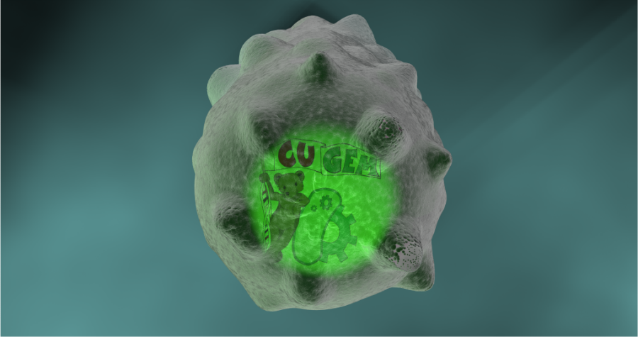
- Light Source
- Show green color and purpose without being too intrusive.
Components Development Summary
- Cell Wall
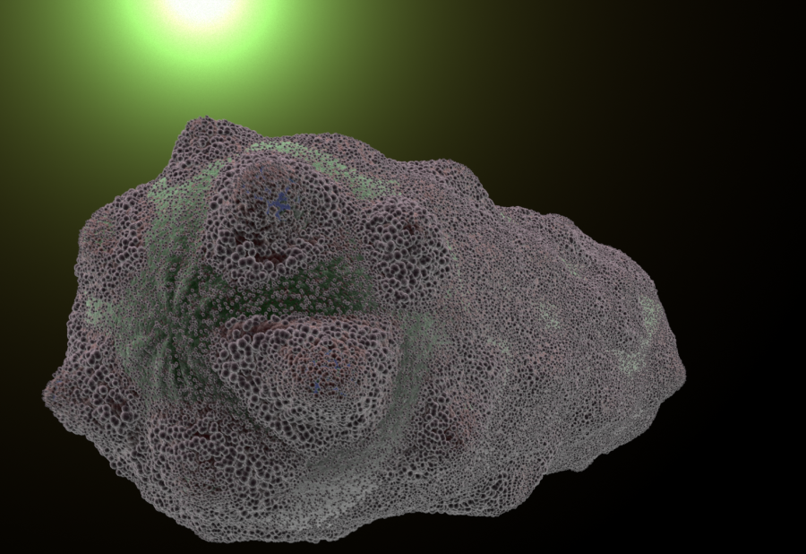
- Created Mesh of E. coli. Sculpted it with a mesh of protoplasm underneath to maintain consistency in cell wall spacing. Sculpted morph maps to show wall "gaining holes." At this point in time, skills were very limited. No viable nice way to explode this.
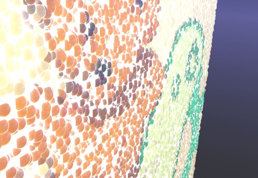
- Replicator Coating - an actual cell wall. Designed by coating the protoplasm mesh with thousands of individual "platelet" meshes. With a surface generator and vertex map deformers, was able to manipulate it very nicely. Individual platelets also helped give E. coli more life.
- Protoplasm
- Created mesh of protoplasm underneath cell wall. Worked on appearance. Team leader requested "wispy" protoplasm. Although cell wall mesh was scratched, protoplasm mesh was very usable and underwent vertex map deformation in sync with its replicator cell wall shell.
- Surface Proteins
- Modeled a stand-in protein model in modo. Textured with a modified version of Yazan Malkosh's electron microscope texture. Team liked appearance.
- Coating Pattern
- Used vertex map deformers. Took several attempts to get right.
- Explosion Technique:
- Experimented with "gooey" and "hard" explosions for meshes. Not satisfied.
- Experimented with dynamics package. Far too resource intensive. Decided replicator techniques would be more effective.
- Resultantly used a combination of vertex map deformers and morph maps on all replicators and meshes. Several renditions --> Settled on "pop and expand" rendition.
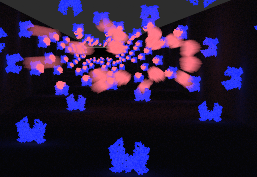
- Beams of Light:
- Started using volumetric direction light sources. Didn't quite like the result. Experimented. Settled on volumetric point light source with light shift over time. Also gave the scene an effective "Sun"
Summary of Scene Progression
For the lysis scene, Modo skill set was still being developed. As a result, scene involved a good amount of trial and error with various scene rewrites.
- Pure Mesh Attempt.(06/26/2011 - 07/03/2011)
Looked very nice in stills. Scratched before significantly animated.
- 1st Draft - Concept visualization. (07/03/2011 - 07/10/2011)
Created two animation models for the major elements of the scene.
The first illustrated replicator offset explosions with holes in the cell wall. Positive Reception.
The second showcased surface protein patterns and deformations. Needed some critiquing - rings vs. globs.
- 2nd Draft - Flagella, Scale Up + Replicator explosion. (07/10/2011 - 07/11/2011)
Resulted from an experiment with flagella. Protoplasm mesh is coated with a cell wall replicator that does not deform. Scaling up the protoplasm resulted in the expansion and "disintegration" of the cell wall. Flagella was dynamically parented to protoplasm for smooth motion weight gradient. Met with positive reception, but team decided to cut flagella for next rendition0. Rendition also did not include surface proteins.
- 3rd Draft - 1st Full Assembly. (07/11/2011 - 07/15/2011)
Rebuilt scene using feedback and lessons from previous renditions.
- Visual Overhaul. Used edited version of Yazan Malkosh's electron microscope texture for cell wall platelets
- Updated and refined vertex map deformers.
- Refined explosion to include organic scaling of bacteria and post-explosion swirling of cell wall platelets and expansion of protoplasm.
Mostly positive reception. Team leader wanted more prominent "spikey" deformations at holes, and more of a burst explosion. Consensus - camera work made bacteria appear to shrink. Also needed to implement surface protein layers.
- 4th Draft - 2nd Full Assembly (07/15/2011 - 07/20/2011)
Refined scene
- Rebuilt and implemented surface protein vertex map deformers. Protein visuals updated.
- Implemented changes to hole deformations. More of a spikey look.
- Modified camera work.
- Refined Explosion - more of a solid pop and burst.
- Updated green light source
- Changed protoplasm to a less resource intensive material.
Positive reception. Results most satisfactory. Comments that holes were too spikey.
At present, will correct if have time
- Experimental Methods (08/08/2011 - present)
- Never implemented in-scene. Post-2nd full assembly, developed a technique for nicer cell wall disintegration around holes. Technique would involve a complete rebuild. If time, plan to implement.
Reception and Review: Satisfactory. Potentially scene could use some slight improvements - namely "spikes" and end protoplasm appearance. Otherwise, current result is nicely polished.