Team:Washington/Magnetosomes/Magnet Results
From 2011.igem.org
| Line 3: | Line 3: | ||
<center><big><big><big><big>'''Magnetosome Toolkit: Results Summary'''</big></big></big></big></center><br><br> | <center><big><big><big><big>'''Magnetosome Toolkit: Results Summary'''</big></big></big></big></center><br><br> | ||
| + | |||
| + | == Magnetosome gene-protein Fusions== | ||
| + | |||
| + | Using our two genes of interest, we created C-terminal sfGFP fusions so we could track the localization of each gene separately within ''E.coli.'' | ||
| + | |||
| + | [[File:Igem2011_mamK_and_I.png|700px|center]] | ||
| + | |||
| + | The results we obtained with our sfGFP fusions inside ''E.coli'' were comparable to those done through other studies in the host organism ''Magnetospirillum magneticum''. In the image of mamK, a filament is seen running through the length of many bacteria. For mamI, the gene product is seen to fluoresce around the cell membrane of the bacteria but mostly concentrated at the ends. Similarly, the graph shows that as the arrow cross the cell membrane, the fluorescent peaks are at a maximum, and through the center of the cell, the level of fluorescence decreases. | ||
| + | |||
| + | ==Construction of the R5 region of the Magnetosome Island in ''E.coli''== | ||
| + | |||
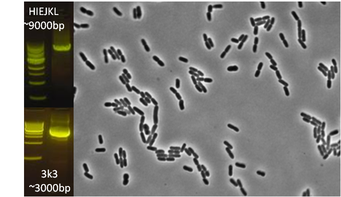
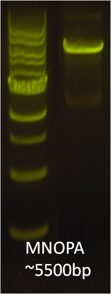
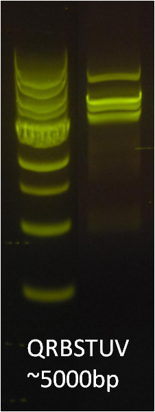
| + | After identifying that the construction of the scaffold had worked, we proceeded to work on the final assembly in three parts: mamHIEJKK, mamMNOPA, and mamQRBSTUV. The PCR products of the first, and the third part of the assembly are shown below. Both fragments of the assembly have been partially sequenced confirmed, and we are currently working on designing primers to fill in the gap sequences. Despite these gaps, when this samples were imaged, filaments in the first part (mamHIEJKL)were still apparent. <br/> | ||
| + | <center>[[File:Washington_iGEM2011_magentosome_HIEJKL3k3.png|400px|middle]][[File:Washington_iGEM2011_magentosome_MNOPA.png|100px|middle]][[File:Washington_iGEM2011_magentosome_QRBSTUV.png|100px|middle]] | ||
| + | </center> | ||
Revision as of 05:42, 23 September 2011
Magnetosome gene-protein Fusions
Using our two genes of interest, we created C-terminal sfGFP fusions so we could track the localization of each gene separately within E.coli.
The results we obtained with our sfGFP fusions inside E.coli were comparable to those done through other studies in the host organism Magnetospirillum magneticum. In the image of mamK, a filament is seen running through the length of many bacteria. For mamI, the gene product is seen to fluoresce around the cell membrane of the bacteria but mostly concentrated at the ends. Similarly, the graph shows that as the arrow cross the cell membrane, the fluorescent peaks are at a maximum, and through the center of the cell, the level of fluorescence decreases.
Construction of the R5 region of the Magnetosome Island in E.coli
After identifying that the construction of the scaffold had worked, we proceeded to work on the final assembly in three parts: mamHIEJKK, mamMNOPA, and mamQRBSTUV. The PCR products of the first, and the third part of the assembly are shown below. Both fragments of the assembly have been partially sequenced confirmed, and we are currently working on designing primers to fill in the gap sequences. Despite these gaps, when this samples were imaged, filaments in the first part (mamHIEJKL)were still apparent.


 "
"


