Team:BU Wellesley Software/Notebook/MargauxNotebook
From 2011.igem.org
(→Week of 6/12 to 6/18) |
(→Week of 6/12 to 6/18) |
||
| Line 7: | Line 7: | ||
!colspan="6"|Protocol List | !colspan="6"|Protocol List | ||
|- | |- | ||
| - | + | |Ligation || [[LigationProtocol.jpg]] | |
| - | | | + | |Restriction Digest|| [[Restriction Digest.jpg]] |
| - | | | + | |Transformation|| [[Transformation Digest.jpg]] |
|} | |} | ||
| + | |||
Revision as of 19:56, 25 June 2011
| Protocol List | |||||
|---|---|---|---|---|---|
| Ligation | LigationProtocol.jpg | Restriction Digest | Restriction Digest.jpg | Transformation | Transformation Digest.jpg |
Contents |
6/1/11-6/6/11:BU-WELLESLEY TEAM BOOT CAMP
The boot camp gave me an opportunity to meet the Wellesley students and learn more about the computational/software side of the team.
Biology:
- Reviewed a lot of terms and concepts that I had not seen in two years
- How to use restriction enzymes to cut and place different parts of the biobrick
- Saw the lab and practiced using a pipette, running a gel, etc.
- Watched an interesting background talk on tuberculosis and how we will assist in the efforts to understanding it
- TB transitions from an active to a latent state
- Hard to treat because the bacteria deceives the body's own immune system by covering itself in lipids
Computational:
- Difficulties in setting up the logic behind using inverstases
- Built a "hello world" application on Clotho as an introduction as to how create new applications
- Explored Clotho and its pros and cons
To practice techniques, we performed mini preps on a variety of previously transformed biobricks. Mine was Lac Z. The quantification of DNA isolated was as follow:
- Lacz.1 105.4 ng/microL
- Lacz.2 52.2 ng/microL
- Lacz.3 28.8 ng/microL
These sample were then run on the a gel next to a ladder and the DNA was confirmed to be present.
In the lab meeting, we began looking at the parts and the part registry page to build up some new plasmids with different parts. I worked with Shannon to find three RBS from the Anderson Collection. We chose the following based on their antibiotic resistance and strength:
1.Bba_J61101
 -On both 2010/2011 plates  (2010 Distribution Plate 1-Well 5L)
 -Resistance A
 -11.9% (strength)
2.Bba_J61127
 -On both 2010/2011 plates (2010 Distribution Plate 1-Well 11N)
 -Resistance A
 -6.5% (strength)
3.Bba_J61100
 -On both 2010/2011 plates (2010 Distribution Plate 1-Well 5J)
 -Resistance A
 -4.75% (strength)
We then plated them and set them aside to grow.
We had the weekly lab meeting and discussed the work flow to build constructs. We also noted that our bacteria with the three different RBS had grown with two different colored colonies. We removed a piece of each colony in each sample (3 samples, 2 colonies each, 6 tubes) and did a prep for a plasmid prep.
Performed a plasmid prep on all six samples. The bacteria that grew during the prep for the plasmid prep showed stringy lifeforms and bacteria in forms that we were not expecting. DNA quantification was low, and the gel electrophoresis showed nothing notable. We then checked the bacteria samples under a light microscope with a gram stain and saw that while the Ecoli we intended to be there was there, there were also other types of bacteria that were contaminating the sample.
DNA Quantification
- Bba_J61101 A 5.2 ng/microL
- Bba_J61101 B 11.2 ng/microL
- Bba_J61100 A 2.6 ng/microL
- Bba_J61100 B 6.5 ng/microL
- Bba_J61127 A 11.4 ng/microL
- Bba_J61127 B 8.4 ng/microL
Autoclaved all our tips to make sure that they were not the source of contamination. Researched about how to build
the constructs we are hoping to make.
Week of 6/12 to 6/18
At the end of the last week, we decided there were two main goals for this week. One was to figure out what was causing the contamination and preventing the transformation of the promoters and ribosome binding sites (RBS). The other was to attempt to piece together the green fluorescent protein (GFP) with the terminator. Both of these blocks had been previously transformed and were currently available to us as plasmids. We split the team up to work on these goals. Alberto and I were predominantly in charge of the first attempt of combining the GFP and terminator, which is described below. A lesser third goal was to prepare plasmids for learning about the QIA cube, a technology that would take care of such techniques as plasmid mini-preps and gel extraction for us.
- Combining GFP and Terminator
As we had the plasmids already available, our first step was to use restriction digest enzymes to cut out appropriate pieces.
GFP Gene: Bba_J52028
Concentration:53.4 ng/microL
Sites Cut: EcoRI, SpeI
Terminator: Gene Bba_B0015
Concentration: 48.5 ng/microL
Sites Cut: EcorI, XbaI
We cut the plasmids at the sites noted according to the following protocol: media:RestrictionDIgest61311_MC.jpg . As the GFP gene was large enough to isolate from the plasmid, we cut it completely out of the plasmid. We only made an incision in the terminator plasmid at the EcoRI and XbalI sites in order to make room for the GFP gene. Subsequent to the restriction digest, we ran the pieces out on a gel. The result of the gel electrophoresis is the following:
The first column is the DNA ladder. The next column is the cut plasmid containing the terminator and the third one is the intact plasmid for comparison. The placement of both was to be expected as the terminator plasmid without a cut is able to supercoil. Thus, it can proceed further down the gel then the cut plasmid. The next two pieces are the cut GFP plasmid and the intact GFP plasmid. The cut one shows two pieces: the higher up piece is the backbone and the cut GFP is the lower one. The GFP plasmid that is intact shows expected placement.
We then proceeded to extract the DNA from the gel. The blocks we desired to remove were the GFP gene and the terminator plasmid. After running the gel extraction protocol (media:gelextractionprotocol61311_MC.jpg) on these blocks we used the Nano-Drop (media:NanoDrop Protocol_MC.jpg) to quantify how much DNA we removed.
GFP    2.4 ng/microL
Terminator 2.7 ng/microL
We were hoping for large amounts, but proceed onto ligating these pieces together anyways. After running the ligation protocol on the two pieces, we proceeded to use the new, combined plasmid in transformation. The goal of this was to acquire Ecoli who would pick up the plasmid. We then performed a mini-prep on these transformations and isolated the plasmids. We then quantified the DNA present and we were pleased that the numbers had increased.
These same steps(from restriction digest to mini-prep) were repeated in order to accumulate as much GFP+Terminator Composite as possible. The repeats were run by Kyle and Alberto.
- QIA Cube
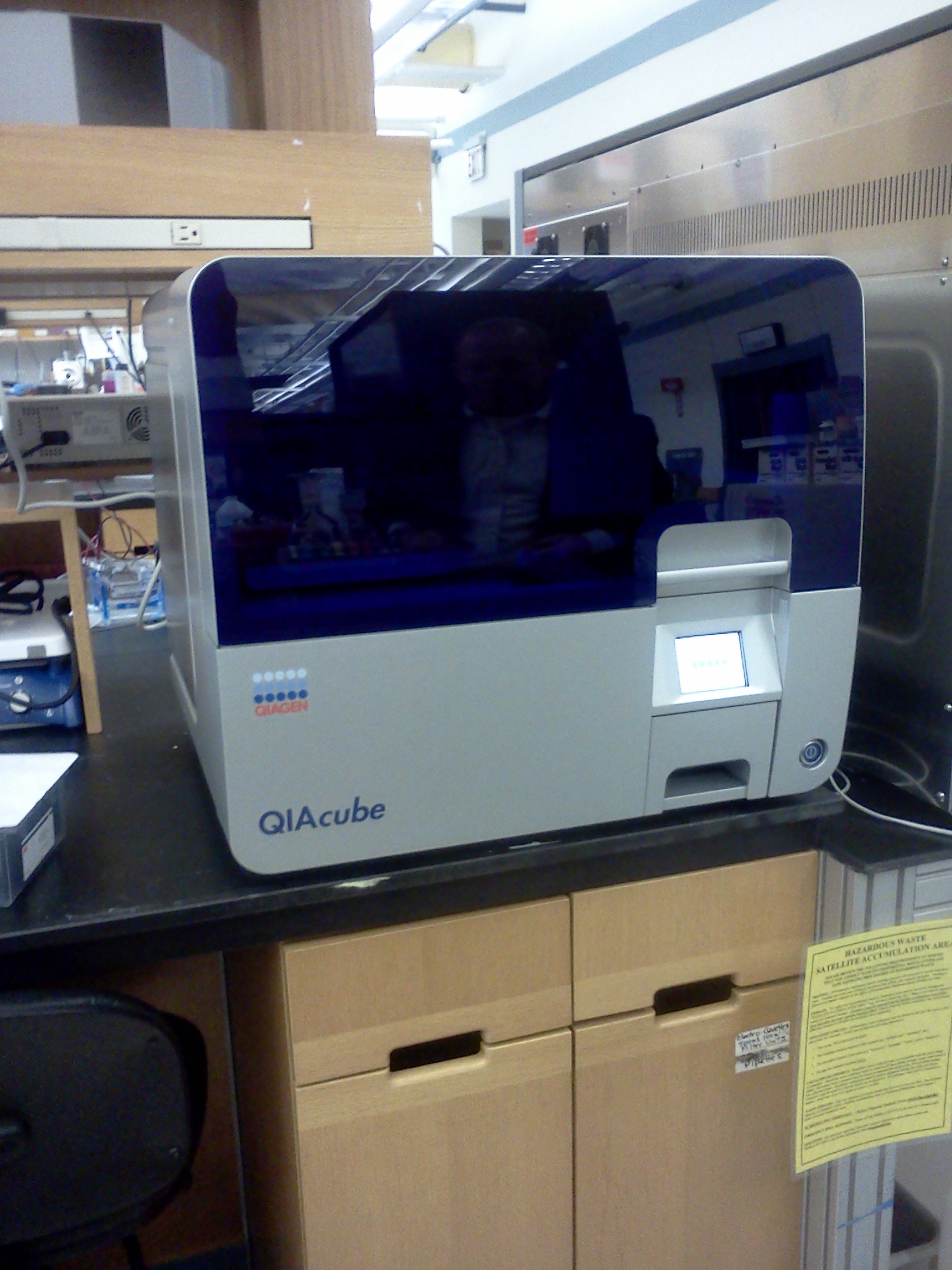
On Tuesday we began a week-long trial of the QIA cube. It is shown below.
We place the tray shown in purple into the machine with the buffers and reagents needed for whatever protocol we want to run (mini-prep,gel extraction). The samples and tips are also put in along with elution columns. It was fairly easy to learn how to use. There are protocol sheets that one can download offline that detail exactly where to place the reagents and buffers. We used it for both gel extraction and mini preps throughout the week. In a lab situation such as ours it was handy up until a point. It did not expedite the procedures, but just freed us up to work on something else. Given the nature of our problems with contamination, we all had to wait on the same procedure in order to proceed. However, if we all had different tasks and were not trying to isolate a problem it would have been very useful.
- RBS and Promoters
After autoclaving the tips on Friday, we proceeded to re-transform the RBS and promoters we were trying to transform the week before. The growth of the transformed bacteria ranged from non-existent to irregular. A few appeared normal, but enough were wrong that we were convinced we had found the problem. We then made new plates with ampicilin. Happily, the new 2011 plates of DNA had come in the same day. Therefore, once the plates were made, we transformed the new DNA. We then completed the plasmid preps the next day and on Friday performed the minipreps. We will move forward with these samples over the next week and hopefully begin constructing full plasmids with the GFP and Terminator Composite we finished making.
Week of 6/19 to 6/25
The overall goal for this week was to start combining parts now that we were able to grow them without contamination. There were a lot of meetings this week between the different teams to help keep us on track.
- Restriction Digest
There were three parts I originally looked to combine this week:
 Bba_E0430
 * contains a composite part of a RBS, YFP, and terminator
 * RBS-B0034; EYFP-E0030; terminator-B0010+B0012;
 Bba_E0240
 * contains a composite part of a RBS, GFP, and terminator
 * RBS-B0032; GFP-E0040; terminator-B0010+B0012;
 Bba_R2000
 * Promoter that has been noted to work well with Bba_E0240
The goal was to then ligate the promoter with both YFPc and GFPc. However, I cut both wrong. Logically, my cuts would have worked but not realistically. I attempted to cut out the promoter and it was too small. The gel came out looking odd and I finally corrected the cuts (media:RD62211_MC.jpg) and they came out looking normal.
- Devices
We were given a goal of coming up with four devices of the following set-up:
Each gene would correspond to a differently colored fluorescent protein. We decided to split up the work to finish making the four devices. I chose to work on making an RFP composite from a variety of promoters we had transformed that had given us pink/red cells.
I used the promoters:
 Bba_J23100
 * has an RBS, RFP, and terminator
 Bba_J23101
 * has an RBS, RFP, and terminator
I cut out, using restriction digest (media:RD62311_MC.jpg), a piece that contained the RBS, RFP, and terminator. I ran them on a gel and the pieces were collected. Their DNA was quantified as
- Bba_J23100.1 1.1 ng/microL
- Bba_J23100.2 2.4 ng/microL
- Bba_J23101.1 16.9 ng/microL
- Bba_J23101.2 11.3 ng/microL
I used the two available cut promoters:
 * Bba_I14033
   Cuts: SpeI, PstI

 * Bba_I13453
   Cuts: SpeI, PstI
to ligate with the RFPc. Their quantification values are noted in this protocol (media:LigationProtocol62311_MC.jpg) along with the values used in the ligation. Transformation was completed with these new plasmids (media:Trans62311_MC.jpg) and nothing grew. My partners had attempted earlier to combine a GFP composite and a YFP composite with promoters and had the same result. Our tendency is to blame our ligation protocol, and are taking steps to correct it.
- Steps Taken
One thing that stood out to me was the low concentration of my parts used in ligation. To acquire a higher concentration,I ran a restriction digest (media:RD62511_MC.jpg) on the following:
BBa_J23101 Bba_J23100 BBa_R2000 Bba_I14033
These were then loaded into a gel and ran.
 "
"


