Team:EPF-Lausanne/Notebook/June2011
From 2011.igem.org
(→Monday, 27 June 2011) |
(→Monday, 27 June 2011) |
||
| Line 187: | Line 187: | ||
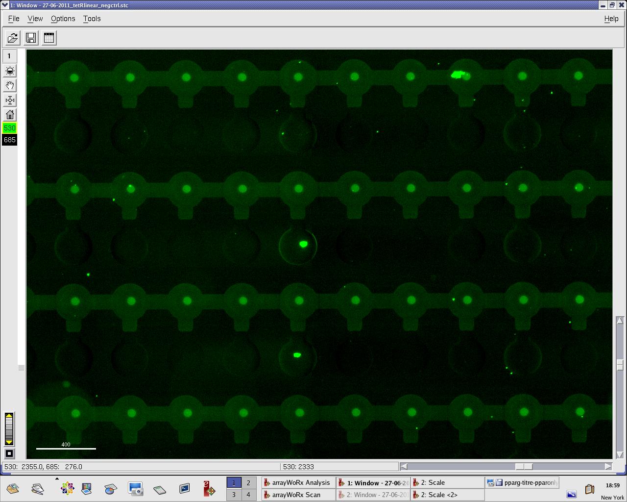
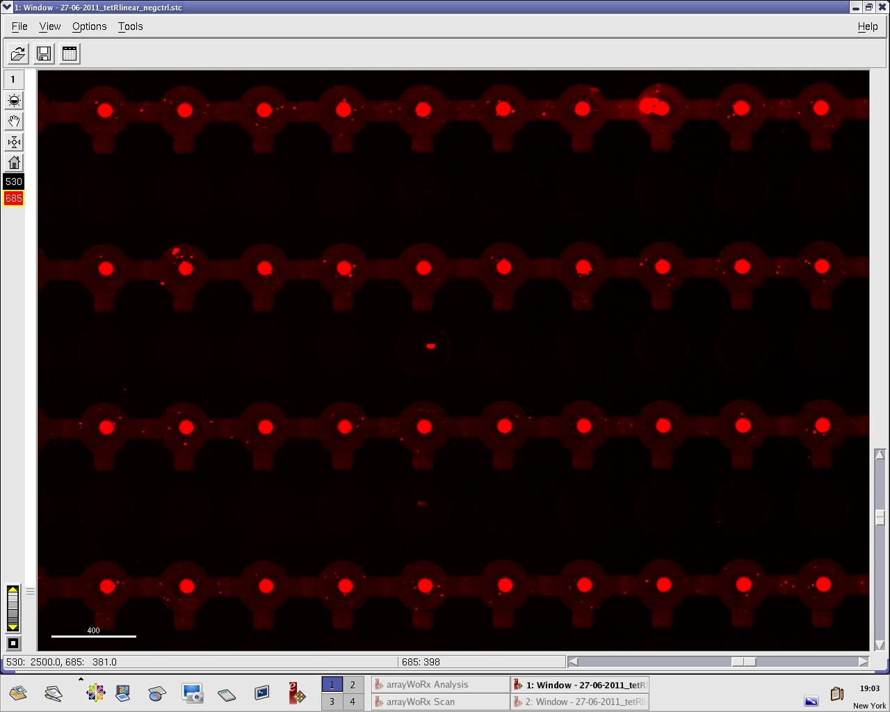
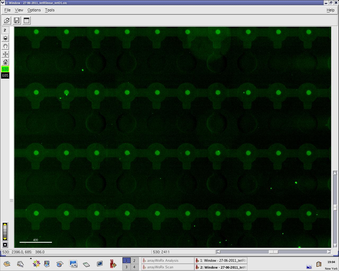
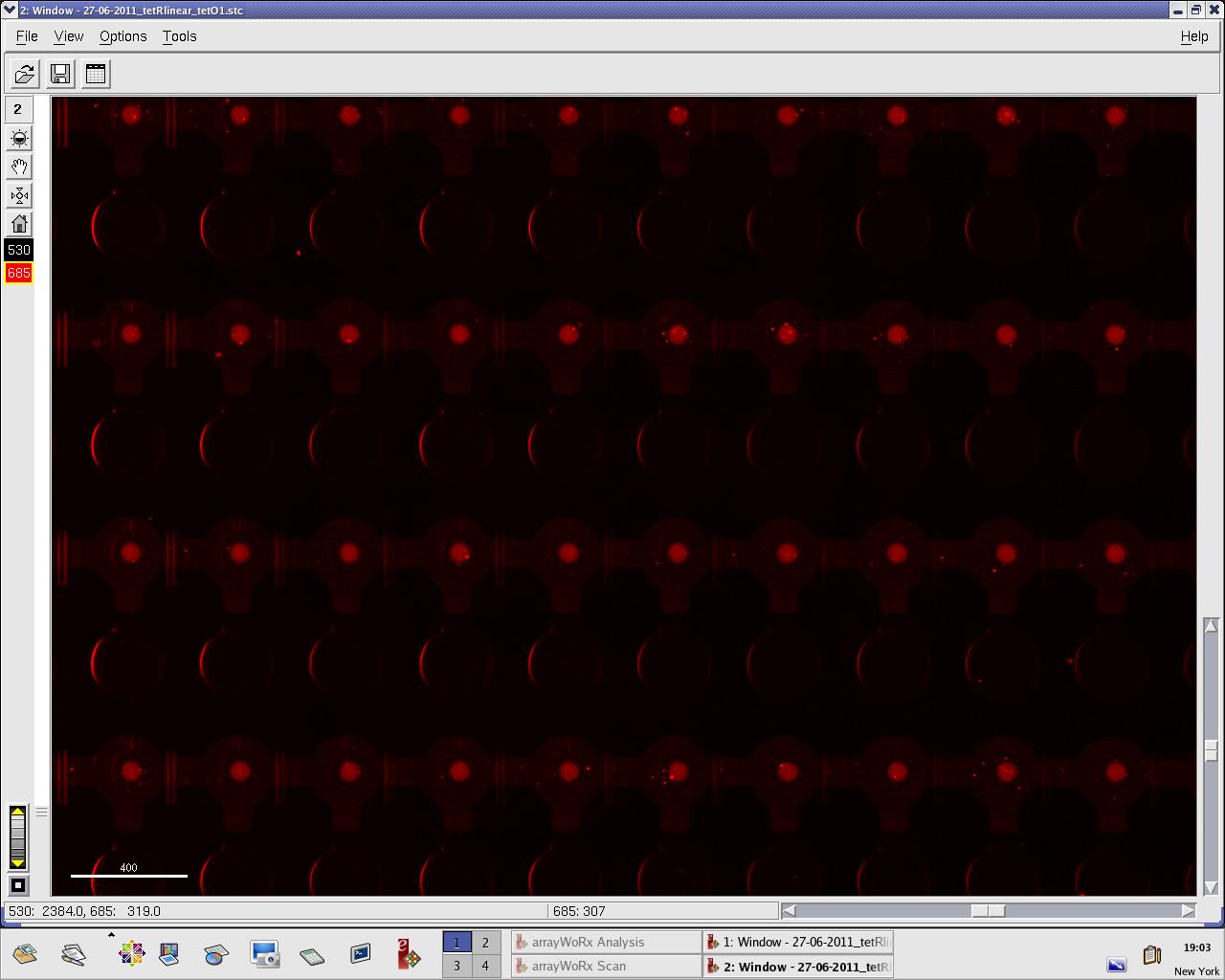
Images: Green fluorescence comes from TetR (labeled Lysines) and red fluorescence comes from DNA (cy-5 labeled). We can clearly see that the protein amount seems to be the same, whereas the DNA fluorescence is decreased. | Images: Green fluorescence comes from TetR (labeled Lysines) and red fluorescence comes from DNA (cy-5 labeled). We can clearly see that the protein amount seems to be the same, whereas the DNA fluorescence is decreased. | ||
| - | [[File:EPFL_27-06-tetR_negctrl_A488.jpg| | + | [[File:EPFL_27-06-tetR_negctrl_A488.jpg|300px|TetR fluorescence during random DNA experiment]] |
| - | [[File:EPFL_27-06-tetR_negctrl_cy5.jpg| | + | [[File:EPFL_27-06-tetR_negctrl_cy5.jpg|300px|DNA fluorescence during random DNA experiment]] |
| - | [[File:EPFL_27-06-tetR_tetO1_A488.jpg| | + | [[File:EPFL_27-06-tetR_tetO1_A488.jpg|300px|TetR fluorescence during target DNA experiment]] |
| - | [[File:EPFL_27-06-tetR_tetO1_cy5.jpg| | + | [[File:EPFL_27-06-tetR_tetO1_cy5.jpg|300px|DNA fluorescence during target DNA experiment]] |
Perhaps the ITT doesn't yield a functional TetR, or we didn't have the optimal concentration of binding DNA (too much nonspecific interactions?). We'll definitely continue the MITOMI practise to find out. | Perhaps the ITT doesn't yield a functional TetR, or we didn't have the optimal concentration of binding DNA (too much nonspecific interactions?). We'll definitely continue the MITOMI practise to find out. | ||
{{:Team:EPF-Lausanne/Templates/Footer}} | {{:Team:EPF-Lausanne/Templates/Footer}} | ||
Revision as of 08:05, 30 June 2011
Notebook: June 2011
Contents |
Friday, 3 June 2011
Lysis device sequences were received from Microsynth. Only two reactions worked: those containing primers 1289F and 1333R.
Output (For the reverse primers, the reverse complement sequence is shown):
  >1556291 30F
  NNNNN

  >1556292 440F
  NNNNN

  >1556293 947F
  NNNNN

  >1556294 1289F
  NNNCTTCGGNGGGCCTTTCTGCGTTNNNATACTANTAGCGGCCGCTGCAGTCCGGCAAAAAAGGGCAAGGTGTCACCACCCTGCCCTTTTTCTTTAAAAC
  CGAAAAGATTACTTCGCGTTATGCAGGCTTCCTCGCTCACTGACTCGCTGCGCTCGGTCGTTCGGCTGCGGCGAGCGGTATCAGCTCACTCAAAGGCGGT
  AATACGGTTATCCACAGAATCAGGGGATAACGCAGGAAAGAACATGTGAGCAAAAGGCCAGCAAAAGGCCAGGAACCGTAAAAAGGCCGCGTTGCTGGCG
  TTTTTCCACAGGCTCCGCCCCCCTGACGAGCATCACAAAAATCGACGCTCAAGTCAGAGGTGGCGAAACCCGACAGGACTATAAAGATACCAGGCGTTTC
  CCCCTGGAAGCTCCCTCGTGCGCTCTCCTGTTCCGACCCTGCCGCTTACCGGATACCTGTCCGCCTTTCTCCCTTCGGGAAGCGTGGCGCTTTCTCATAG
  CTCACGCTGTAGGTATCTCAGTTCGGTGTAGGTCGTTCGCTCCAAGCTGGGCTGTGTGCACGAACCCCCCGTTCAGCCCGACCGCTGCGCCTTATCCGGT
  AACTATCGTCTTGAGTCCAACCCGGTAAGACACGACTTATCGCCACTGGCAGCAGCCACTGGTAACAGGATTAGCAGAGCGAGGTATGTAGGCGGTGCTA
  CAGAGTTCTTGAAGTGGTGGCCTAACTACGGCTACACTAGAAGAACAGTATTTGGTATCTGCGCTCTGCTGAAGCCAGTTACCTTCGGAANAAGAGTTGG
  TAGCTCTTGATCCGGCAAACAAACCACCGCTGGTAGCGGTGGTTTTTTTGTTTGCAAGCAGCAGATTACGCGCAGAAAANAAGGATCTCAAGAAGATCCT
  TTGATCTTTTCTACGGGGTCTGACGCTCAGTGGAACGAANACTCACGTTAAGGGATTTTGGTCATGAGATTATCAAAAAGGATCTTCACCTAGATCCTTT
  TAAATTAAAAATGAAGTTTTANATCAATCTAAAGTATATATGANTAAACTTGGTCTGANAGCTCGAGATTCTCATGTTTGANAGCTTATCATCGATAAGC
  TTTAATGNGGTAGTTTATCNNAGTTAAATTGCTAACGCAGTCAGGNNCCGNN

  >1556295 816R
  NNNNN

  >1556296 1333R
  TNCNTTNCTAGAGGGNCAAGCAATGCTTGCCTTGAATANNAACTTTTGAATANNGATTCAGGAGGTACTAGAATGGCTTCCAAGGTGTACGACCCCGAGC
  AACGCAAACGCATGATCACTGGGCCTCAGTGGTGGGCTCGCTGCAAGCAAATGAACGTGCTGGACTCCTTCATCAACTACTATGATTCCGAGAAGCACGC
  CGAGAACGCCGTGATTTTTNTGCATGGTAACGCTGCCTCCAGCTACCTGTGGAGGCACGTCGTGCCTCACATCGAGCCCGTGGCTAGATGCATCATCCCT
  GATCTGATCGGAATGGGTAAGTCCGGCAAGAGCGGGAATGGCTCATATCGCCTCCTGGATCACTACAAGTACCTCACCGCTTGGTTCGAGCTGCTGAACC
  TTCCAAAGAAAATCATCTTTGTGGGCCACGACTGGGGGGCTTGTCTGGCCTTTCACTACTCCTACGAGCACCAAGACAAGATCAAGGCCATCGTCCATGC
  TGAGAGTGTCGTGGACGTGATCGAGTCCTGGGACGAGTGGCCTGACATCGAGGAGGATATCGCCCTGATCAAGAGCGAAGAGGGCGAGAAAATGGTGCTT
  GAGAATAACTTCTTCGTCGAGACCATGCTCCCAAGCAAGATCATGCGGAAACTGGAGCCTGAGGAGTTCGCTGCCTACCTGGAGCCATTCAAGGAGAAGG
  GCGAGGTTAGACGGCCTACCCTCTCCTGGCCTCGCGAGATCCCTCTCGTTAAGGGAGGCAAGCCCGACGTCGTCCAGATTGTCCGCAACTACAACGCCTA
  CCTTCGGGCCAGCGACGATCTGCCTAAGATGTTCATCGAGTCCGACCCTGGGTTCTTTTCCAACGCTATTGTCGAGGGAGCTAAGAAGTTCCCTAACACC
  GAGTTCGTGAAGGTGAAGGGCCTCCACTTCAGCCAGGAGGACGCTCCAGATGAAATGGGTAAGTACATCAAGAGCTTCGTGGAGCGCGTGCTGAAGANNN
  GAGCAGTAAACTAGAGCCAGGCATCAAATAAAACGAAAGGNTCAGTCGAAAGACTGGGCCTTTCGTTTNNNN

  >1556297 1734R
  NNNNN
Monday, 6 June 2011
Irina and Nadine selected the 2 plasmid backbones we will use: J23019 and J61002 (check the parts registry). They have 2 different origin of replication sites and should both have 15-20 copy numbers. Interesting also, they both contain RFP and pTet. We already have them in the freezers, now let's focus on primers design!
Wednesday, 8 June 2011
Lilia and Douglas miniprep'ed the cultures containing the lysis cassette, and sent the plasmids for sequencing.
Three colonies were miniprep'ed: two containing the T4 variant, one containing the lambda variant. This yielded solutions with plasmid concentrations of 221.9 ng/µl for T4 colony 1, 225.5 ng/µl for T4 colony 2, and 57.0 ng/µl for lambda colony. Only T4 colony 1 was sent for sequencing, using the primer solutions prepared on 31 May 2011. Results should be in on Friday.
Friday, 10 June 2011
Sequencing results from Microsynth for the lysis cassette (Biobrick K112808) have arrived. The sequence is entirely covered by the seven reactions, and all but a few bases match. As far as I can tell, this biobrick is valid! (Doug)
The results of a blast, individually querying each of the sequences against the official K112808 sequence are available here: BLAST results.
Sequencing results, for each primer (again, reverse complements shown here for the reverse primers --- those denoted by an "R"):
   >1558332 30F
   NGNNNTCATTTGGTGTTCAGATCGCTTGTTCAAGATAACGCTACCGGGAAGGTTCTTGCTTCCCGGGTAGCTGTCGTAATTCTTTTGTTTATAATGGCGA
   TTGTTTGGTATAGGGGAGATAGTTTCTTTGAGTACTATAAGCAATCAAAGTATGAAACATACAGTGAAATTATTGAAAAGGAAAGAACTGCACGCTTTGA
   ATCTGTCGCCCTGGAACAACTCCAGATAGTTCATATATCATCTGAGGCAGACTTTAGTGCGGTGTATTCTTTCCGCCCTAAAAACTTAAACTATTTTGTT
   GATATTATAGCATACGAAGGAAAATTACCTTCAACAATAAGTGAAAAATCACTTGGAGGATATCCTGTTGATAAAACTATGGATGAATATACAGTTCATT
   TAAATGGACGTCATTATTATTCCAACTCAAAATTTGCTTTTTTACCAACTAAAAAGCCTACTCCCGAAATAAACTACATGTACAGTTGTCCATATTTTAA
   TTTGGATAATATCTATGCTGGAACGATAACCATGTACTGGTATAGAAATGATCATATAAGTAATGACCGCCTTGAATCAATATGTGCTCAGGCGGCCAGA
   ATATTAGGAAGGGCTAAATAATTAACTAGAATACTTAGGAGGTATTATGAATATATTTGAAATGTTACGTATAGATGAAGGTCTTAGACTTAAAATCTAT
   AAAGACACAGAAGGCTATTACACTATTGGCATCGGTCATTTGCTTACAAAAAGTCCATCACTTAATGCTGCTAAATCTGAATTAGATAAAGCTATTGGGC
   GTAATTGCAATGGTGTAATTACAAAAGATGAGGCTGAAAAACTCTTTAATCAGGATGTTGATGCTGCTGTTCGCGGAATCCTGAGAAATGCTAAATTAAA
   ACCGGTTTATGATTCTCTTGATGCGGTTCGTCGCTGTGCATTGATTAATATGGTTTTCCAAATGGGANAAACCGGTGTGGCAGGATTTACTAACTCTTTA
   CGTATGCTTCAACAAAAACGCTGGGATGAAGCAGCAGTTAACTTAGCTAANAGTAGATGGNNTAATCAAACNCCTAATCNNNNAAAACGAGTCATTACAA
   CGTTTANAACTGGNNN

   >1558333 440F
   NNNATTCCNACTCAAATTTGCTTTTTTACCAACTAAAAAGCCTACTCCCGAAATAAACTACATGTACAGTTGTCCATATTTTAATTTGGATAATATCTAT
   GCTGGAACGATAACCATGTACTGGTATAGAAATGATCATATAAGTAATGACCGCCTTGAATCAATATGTGCTCAGGCGGCCAGAATATTAGGAAGGGCTA
   AATAATTAACTAGAATACTTAGGAGGTATTATGAATATATTTGAAATGTTACGTATAGATGAAGGTCTTAGACTTAAAATCTATAAAGACACAGAAGGCT
   ATTACACTATTGGCATCGGTCATTTGCTTACAAAAAGTCCATCACTTAATGCTGCTAAATCTGAATTAGATAAAGCTATTGGGCGTAATTGCAATGGTGT
   AATTACAAAAGATGAGGCTGAAAAACTCTTTAATCAGGATGTTGATGCTGCTGTTCGCGGAATCCTGAGAAATGCTAAATTAAAACCGGTTTATGATTCT
   CTTGATGCGGTTCGTCGCTGTGCATTGATTAATATGGTTTTCCAAATGGGAGAAACCGGTGTGGCAGGATTTACTAACTCTTTACGTATGCTTCAACAAA
   AACGCTGGGATGAAGCAGCAGTTAACTTAGCTAAAAGTAGATGGTATAATCAAACACCTAATCGCGCAAAACGAGTCATTACAACGTTTAGAACTGGCAC
   TTGGGACGCGTATAANAATCTATAAAGCACTAGAGCCAGGCATCAAATAAAACGAAAGGCTCAGTCGAAAGACTGGGCCTTTCGTTTTATCTGTTGTTTG
   TCGGTGAACGCTCTCTACTAGAGTCACACTGGCTCACCTTCGGGTGGGCCTTTCTGCGTTTATATACTAGAGTTGACAGCTAGCTCAGTCCTAGGGACTA
   TGCTAGCTACTAGAGATAGGAGGCCTTTATGGCCTTAAAAGCAACAGCACTTTTTGCCATGCTAGGATTGTCATTTGTTTTATCTCCATCGATTGAAGCG
   AATGTCNATCCTCATTTTGATAAATTTATGGAATCTGGNNTTAGGCACGTTTATNTGCTTTTTGAAAANAAAANCGTANAATCNNCTGAACAATTCTATA
   GTTTTANNNNAACGACCTATAAAANTGACCCNNGN

   >1558334 947F
   NNTTGNNGCGGTTCGTCGCTGTGCATTGATTAATATGGTTTTCCAAATGGGAGAAACCGGTGTGGCAGGATTTACTAACTCTTTACGTATGCTTCAACAA
   AAACGCTGGGATGAAGCAGCAGTTAACTTAGCTAAAAGTAGATGGTATAATCAAACACCTAATCGCGCAAAACGAGTCATTACAACGTTTAGAACTGGCA
   CTTGGGACGCGTATAAAAATCTATAAAGCACTAGAGCCAGGCATCAAATAAAACGAAAGGCTCAGTCGAAAGACTGGGCCTTTCGTTTTATCTGTTGTTT
   GTCGGTGAACGCTCTCTACTAGAGTCACACTGGCTCACCTTCGGGTGGGCCTTTCTGCGTTTATATACTAGAGTTGACAGCTAGCTCAGTCCTAGGGACT
   ATGCTAGCTACTAGAGATAGGAGGCCTTTATGGCCTTAAAAGCAACAGCACTTTTTGCCATGCTAGGATTGTCATTTGTTTTATCTCCATCGATTGAAGC
   GAATGTCGATCCTCATTTTGATAAATTTATGGAATCTGGTATTAGGCACGTTTATATGCTTTTTGAAAATAAAAGCGTAGAATCGTCTGAACAATTCTAT
   AGTTTTATGAGAACGACCTATAAAAATGACCCGTGCTCTTCTGATTTTGAATGTATAGAGCGAGGCGCGGAGATGGCACAATCATACGCTAGAATTATGA
   ACATTAAATTGGAGACTGAATGANATACTAGAGCCGGCTTATCGGTCAGTTTCACCTGATTTACGTAAAAACCCGCTTCGGCGGGTTTTTGCTTTTGGAG
   GGGCAGAAAGATGAATGACTGTCCACGACGCTATACCCAAAAGAAATACTAGTCTGCAGGCTTCCTCGCTCACTGACTCGCTGCGCTCGGTCGTTCGGCT
   GCGGCGAGCGGTATCAGCTCACTCAAAGGCGGTAATACGGTTATCCACAGAATCAGGGGATAACGCAGGAAAGAACATGTGAGCAAAAGGCCAGCAAAAG
   GCCAGGAACCGTAAAAAGGCCGCGTTGCTGGCGTTTTTCCNCAGGCTCCGCCCCCCTGACGANCATCNCAAAAATCGACGCTCAAGTCANANGNN

   >1558335 1289F
   NNCTTCGGNGGGCTTTCTGNGTTTATATANTAGAGTTGACAGCTAGCTCAGTCCTAGGGACTATGCTAGCTACTAGAGATAGGAGGCCTTTATGGCCTTA
   AAAGCAACAGCACTTTTTGCCATGCTAGGATTGTCATTTGTTTTATCTCCATCGATTGAAGCGAATGTCGATCCTCATTTTGATAAATTTATGGAATCTG
   GTATTAGGCACGTTTATATGCTTTTTGAAAATAAAAGCGTAGAATCGTCTGAACAATTCTATAGTTTTATGAGAACGACCTATAAAAATGACCCGTGCTC
   TTCTGATTTTGAATGTATAGAGCGAGGCGCGGAGATGGCACAATCATACGCTAGAATTATGAACATTAAATTGGAGACTGAATGAAATACTAGAGCCGGC
   TTATCGGTCAGTTTCACCTGATTTACGTAAAAACCCGCTTCGGCGGGTTTTTGCTTTTGGAGGGGCAGAAAGATGAATGACTGTCCACGACGCTATACCC
   AAAAGAAATACTAGTCTGCAGGCTTCCTCGCTCACTGACTCGCTGCGCTCGGTCGTTCGGCTGCGGCGAGCGGTATCAGCTCACTCAAAGGCGGTAATAC
   GGTTATCCACAGAATCAGGGGATAACGCAGGAAAGAACATGTGAGCAAAAGGCCAGCAAAAGGCCAGGAACCGTAAAAAGGCCGCGTTGCTGGCGTTTTT
   CCACAGGCTCCGCCCCCCTGACGAGCATCACAAAAATCGACGCTCAAGTCAGAGGTGGCGAAACCCGACAGGACTATAAAGATACCAGGCGTTTCCCCCT
   GGAAGCTCCCTCGTGCGCTCTCCTGTTCCGACCCTGCCGCTTACCGGATACCTGTCCGCCTTTCTCCCTTCGGGAAGCGTGGCGCTTTCTCATAGCTCAC
   GCTGTAGGTATCTCAGTTCGGTGTAGGTCGTTCGCTCCAAGCTGGGCTGTGTGCACGAACCCCCCGTTCAGCCCGACCGCTGCGCCTTATCCGGTAACTA
   TCGTCTTGAGTCCAACCCGGTAAGACACGACTTATCGCCACTGGCAGCAGCCACTGGTAANAGGATTAGCANNNNNNGGTATGTAGGNNNNNCTACAGAN
   TTCTTGAAGTGGNGGCCTAACTACGNNTAN

   >1558336 816R
   NTTCAGNNTNTTTTACTTTCACCAGCGTTTNTGGGNGANNAAAAACAGGAAGGCAAAATGCCGCAAAAAAGGGAATAAGGGNGACACGGAAATGTTGAAT
   ACTCATACTCTTCCTTTTTCAATATTATTGAAGCATTTNTCAGGGTTATTGTNTCATGAGCGGATACATATTTGAATGTATTTAGAAAAATAAACAAATA
   GNGGTTCCGCGCACATTTCCCCGAAAAGTGCCACCTGACGTCTAAGAAACCATTATTATCATGACATTAACCTATAAAAATAGGCGTATCACGAGGCAGA
   ATNTCAGATAAAAAAAATCCTTAGCTTTCGCTAAGGATGATTTCTGGAATTCGCGGCCGCTTCTAGACTTAAAAGGAGGGTCTATGGCAGCACCTAGAAT
   ATCATNTTCGCCCTCTGATATTCTATTTGGTGTTCTAGATCGCTTGTTCAAAGATAACGCTACCGGGAAGGTTCTTGCTTCCCGGGTAGCTGTCGTAATT
   CTTTTGTTTATAATGGCGATTGTTTGGTATAGGGGAGATAGTTTCTTTGAGTACTATAAGCAATCAAAGTATGAAACATACAGTGAAATTATTGAAAAGG
   AAAGAACTGCACGCTTTGAATCTGTCGCCCTGGAACAACTCCAGATAGTTCATATATCATCTGAGGCAGACTTTAGTGCGGTGTATTCTTTCCGCCCTAA
   AAACTTAAACTATTTTGTTGATATTATAGCATACGAAGGAAAATTACCTTCAACAATAAGTGAAAAATCACTTGGAGGATATCCTGTTGATAAAACTATG
   GATGAATATACAGTTCATTTAAATGGACGTCATTATTATTCCAACTCAAAATTTGCTTTTTTACCAACTAAAAAGCCTACTCCCGAAATAAACTACATGT
   ACAGTTGTCCATATTTTAATTTGGATAATATCTATGCTGGAACGATAACCATGTACTGGTATAGAAATGATCATATAAGTAATGACCGCCTTGAATCAAT
   ATGTGCTCAGGCGGCCAGAATATTAGGAAGGGCTAAATAATTAACTAGAATACTTAGGAGGTATTATGAATATATTTGAAATGTTACGTATAGATNAAGG
   TCTAGACTAAANNCN

   >1558337 1333R
   CCGGGAAGGTTNTTNNNTCCCGGGTAGNNNNCGTAATNNTTNNNTNNNAATGNNGATTGTTTGTNTAGGGGAGATAGTTTNTTTGAGTACNATAAGCAAT
   CAAAGTNTGAAACATCCAGNGAAATTATTGAAAAGGAAANAACTGCACNCTTTGAATCTNTNGCCCTGGAACAACTCCAGATAGTTCATATNTCATNTGA
   GGCAGACTTTAGTGCGGTGTATTCTNTCCGCCCTAAAAACTTAAACTATTTTGTTGATATTATAGCATACGAAGGAAAATTACCTTCAACAATAAGTGAA
   AAATCACTTGGAGGATATCCTGTTGATAAAACTATGGATGAATATACAGTTCATTTAAATGGACGTCATTATTATTCCAACTCAAAATTTGCTTTTTTAC
   CAACTAAAAAGCCTACTCCCGAAATAAACTACATGTACAGTTGTCCATATTTTAATTTGGATAATATCTATGCTGGAACGATAACCATGTACTGGTATAG
   AAATGATCATATAAGTAATGACCGCCTTGAATCAATATGTGCTCAGGCGGCCAGAATATTAGGAAGGGCTAAATAATTAACTAGAATACTTAGGAGGTAT
   TATGAATATATTTGAAATGTTACGTATAGATGAAGGTCTTAGACTTAAAATCTATAAAGACACAGAAGGCTATTACACTATTGGCATCGGTCATTTGCTT
   ACAAAAAGTCCATCACTTAATGCTGCTAAATCTGAATTAGATAAAGCTATTGGGCGTAATTGCAATGGTGTAATTACAAAAGATGAGGCTGAAAAACTCT
   TTAATCAGGATGTTGATGCTGCTGTTCGCGGAATCCTGAGAAATGCTAAATTAAAACCGGTTTATGATTCTCTTGATGCGGTTCGTCGCTGTGCATTGAT
   TAATATGGTTTTCCAAATGGGAGAAACCGGTGTGGCAGGATTTACTAACTCTTTACGTATGCTTCAACAAAAACGCTGGGATGAAGCAGCAGTTAACTTA
   GCTAAAAGTAGATGGTATAATCAAACACCTAATCGCGCAAAACGAGTCATTACAACGTTTAGAACTGGCACTTGGGACGCGTATAAAAATCTATAAAGCA
   CTAGAGCCAGGCATCAAATAAAACGAAAGGCTCAGTCGAAAGACTGGGCCNTTCGNTTNN

   >1558338 1734R
   TCCAACTCAAAATTTNNTTTNTTNCCAACTAAAAANNCTNNTCCCGAAATAAACTNNANGNNNAGTNNTCCANNTNTTAATTTGGATAANNNNTNNGCTG
   GAACGATAACCATGTACTGGTATAGAAATGATCANNNAAGTAATGACCNCCTTGAATCAATATGTGNTCAGGNGNCCAGAATATTAGGAAGGGCTAAATA
   ATTAACTAGAATACTTAGGAGGTATTATGAATATATTTGAAATGTTACGTATAGATGAAGGTNTTAGACTTAAAATCTATAAAGACACAGAAGGCTATTA
   CACTATTGGCATCGGTCATTTGCTTACAAAAAGTCCATCACTTAATGCTGCTAAATCTGAATTAGATAAAGCTATTGGGCGTAATTGCAATGGTGTAATT
   ACAAAAGATGAGGCTGAAAAACTCTTTAATCAGGATGTTGATGCTGCTGTTCGCGGAATCCTGAGAAATGCTAAATTAAAACCGGTTTATGATTCTCTTG
   ATGCGGTTCGTCGCTGTGCATTGATTAATATGGTTTTCCAAATGGGAGAAACCGGTGTGGCAGGATTTACTAACTCTTTACGTATGCTTCAACAAAAACG
   CTGGGATGAAGCAGCAGTTAACTTAGCTAAAAGTAGATGGTATAATCAAACACCTAATCGCGCAAAACGAGTCATTACAACGTTTAGAACTGGCACTTGG
   GACGCGTATAAAAATCTATAAAGCACTAGAGCCAGGCATCAAATAAAACGAAAGGCTCAGTCGAAAGACTGGGCCTTTCGTTTTATCTGTTGTTTGTCGG
   TGAACGCTCTCTACTAGAGTCACACTGGCTCACCTTCGGGTGGGCCTTTCTGCGTTTATATACTAGAGTTGACAGCTAGCTCAGTCCTAGGGACTATGCT
   AGCTACTAGAGATAGGAGGCCTTTATGGCCTTAAAAGCAACAGCACTTTTTGCCATGCTAGGATTGTCATTTGTTTTATCTCCATCGATTGAAGCGAATG
   TCGATCCTCATTTTGATAAATTTATGGAATCTGGTATTAGGCACGTTTATATGCTTTTTGAAAATAAAAGCGTAGAATCGTCTGAACAATTCTATAGTTT
   TATGAGAACGACCTATAAAAATGACCCGTGCTCTTCTGATTTTGAATGTATAGAGCGAGGCGCGGAGANNNCACAN
Tuesday, 21 June 2011
With help from Henrike, Douglas checked the mistakes in the K112808 (lysis cassette) sequence. On the entire sequence, only 7 errors occurred in several reactions, and are therefore likely to be actual errors in the DNA. They occur in positions (with regard to the original sequence in the registry):
- 70: CTC codon substituded for CTA. Both code for leu, so that position is still valid
The following are missing bases:
- 677 and 684: lie in the junction between sub-bricks K112805 and K112806. Still valid.
- 1199: lies in the junction between K112806 and B0010. Still valid.
- 1387: second base of K112807, in the non-coding region. Still valid.
- 1698: lies in the junction between K112807 and B0010. Still valid.
- 1701: beginning of B0010, in the non-coding region. Still valid.
In conclusion, there is one codon substitution, and six missing bases that all lie within the junction between two bricks or the non-coding region of a brick. Experimentally, the brick does lyse cells in culture. Therefore, the sequence appears valid.
Monday, 27 June 2011
We practised MITOMI with TetR linear template and random DNA sequence + target DNA sequence. We performed ITT (in vitro transcription/translation) on TetR linear template before adding it to the chip. However, the results are disappointing: we have more DNA binding for the random sequence (1000 RFU change) than for the target sequence (500 RFU)...
Images: Green fluorescence comes from TetR (labeled Lysines) and red fluorescence comes from DNA (cy-5 labeled). We can clearly see that the protein amount seems to be the same, whereas the DNA fluorescence is decreased.


Perhaps the ITT doesn't yield a functional TetR, or we didn't have the optimal concentration of binding DNA (too much nonspecific interactions?). We'll definitely continue the MITOMI practise to find out.
 "
"

