Template:Team:HokkaidoU Japan/HumanPractice/Part4/box5
From 2011.igem.org
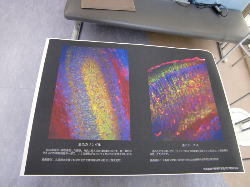
We added a title and an explanation to each picture and printed it out. We also made a video with the moving images provided to show during the event. In this video, we showed stereoscopic image of a cell taken by a confocal laser scanning microscope.
 "
"
