Team:Tsinghua/project
From 2011.igem.org
(→Binding) |
(→Binding) |
||
| Line 28: | Line 28: | ||
OmpA-like transmembrane domain is an evolutionarily conserved domain of outer membrane proteins. This domain consists of an eight-stranded beta barrel. OmpA is the predominant cell surface antigen in enterobacteria found in about 100,000 copies per cell. The expression of OmpA is tightly regulated by a variety of mechanisms. | OmpA-like transmembrane domain is an evolutionarily conserved domain of outer membrane proteins. This domain consists of an eight-stranded beta barrel. OmpA is the predominant cell surface antigen in enterobacteria found in about 100,000 copies per cell. The expression of OmpA is tightly regulated by a variety of mechanisms. | ||
| - | <gallery heights=250px | + | <gallery heights=250px widths=160px perrow=2> |
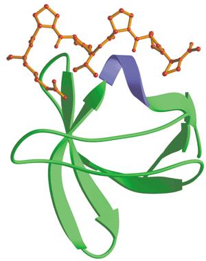
File:SH3_domain.jpg|SH3 binding domain (shown bound to proline-rich peptide) | File:SH3_domain.jpg|SH3 binding domain (shown bound to proline-rich peptide) | ||
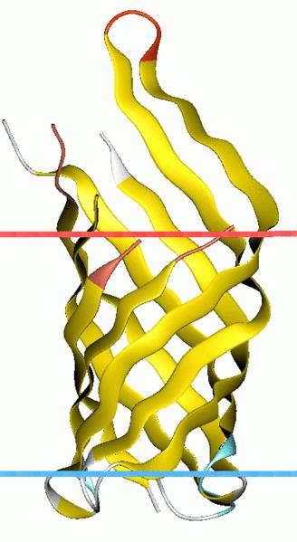
File:OmpA-like_transmembrane_domain.jpg|OmpA transmembrane segment | File:OmpA-like_transmembrane_domain.jpg|OmpA transmembrane segment | ||
Revision as of 05:17, 2 September 2011

E. Colimousine
Overview
This project is destined to generate an E. Coli strain which can move forth and back to transport any desired protein along a gradient. To be specific, the bacteria can bind with the target protein at one place and then, by sensing the gradient of certain amino acid, move to its destination, where it releases the protein with the help from another E. Coli strain. When this is done, it will move back to transport more target protein. To realize our goal, four modules are necessary: binding, releasing, movement and transition.
Binding
Src-homology 3 (SH3) domain has high affinity for proline-rich peptides and together they can form a left-handed poly-Pro type II helix, with the minimal consensus Pro-X-X-Pro. The small size and high affinity are ideal for our carrier design. In our experiment, we used a short peptide with a Kd of 3.67μM (Y. Jacquot, et al., 2007) as the binding motif and planned to transport protein substrates with this peptide sequence.
OmpA-like transmembrane domain is an evolutionarily conserved domain of outer membrane proteins. This domain consists of an eight-stranded beta barrel. OmpA is the predominant cell surface antigen in enterobacteria found in about 100,000 copies per cell. The expression of OmpA is tightly regulated by a variety of mechanisms.
These two domains, as well as the proline-rich peptides are three most important parts if we want to achieve the goal of binding the target protein to the outer membrane of E.coli. First of all, we add proline-rich peptides to the target protein, in this case, mCherry which is easy to detect if the experiment is successful. Then OmpA-SH3 protein is expressed in E.coli and located onto the outer membrane so it can bind with the purified proline-rich containing mCherry protein in the medium.
So, the binding module actually comprises expression of three proteins, namely, OmpA-SH3 protein, which functions as the binding vehicle, proline-rich containing mCherry protein, which functions as the binding substrate and OmpA-mCherry protein, which functions as the positive control.
Releasing
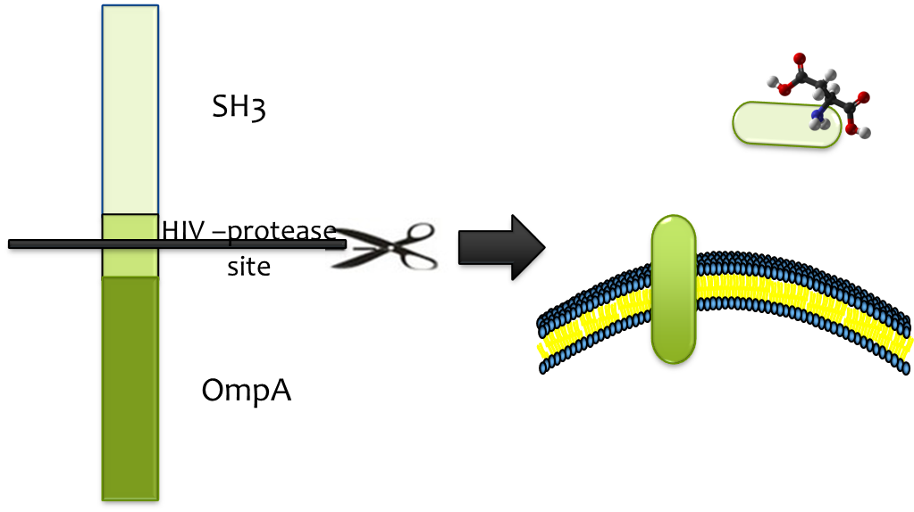
It’s difficult to release the substrate from strong binding, and hence protease is called into play. HIV-protease is readily available and its high efficiency, low molecular weight and high specificity constitutes fine candidate. We only need to add the substrate sequence (HIV-protease site) between OmpA and SH3 sequence and the protease will cut off the binding cassette. In order to segregate the action, we fuse the HIV-protease with OmpA and expressed it in a second E.coli strain which we immobilize at the destination. So, when the E.coli transporter arrives at the destination, the HIV-protease that is located on the outer membrane of the second E.coli strain will help release the substrate (together with SH3 domain) by cut the HIV-protease site.
Movement
Chemotaxis is most frequently used when it comes to the movement of E.coli cells and the regulatory network controlling E.coli chemotaxis is one of the well-studied models of signal transduction. Extracellular signals, aspartate and serine in this case, are sensed by receptors Tar and Tsr respectively. These receptors assemble into large clusters that are predominantly located at the poles of the bacterium. The CheW protein binds to the receptors and acts as a scaffold protein to recruit CheA, whose phosphorylation is stimulated by the receptor. The phosphate is transferred to the cytoplasmic protein CheY, which controls flagellar rotation and in turn, controls the movement of E.coli. Previously, a pair of mutations in CheW* (V108M) and Tsr* (E402A) was shown to produce an orthogonal pair, where the mutated proteins interact with each other but not with wildtype Tsr or CheW. So, this can be used to toggle between activating the serine-responsive Tsr* and aspartate-responsive Tar. When we set up two gradients (serine and aspartate) going in opposite direction, the bacteria can move forth and back.
Transition
We need to shift between two states, the binding state and the releasing state. This molecular device is well-known for its lag in phase change, which is well adapted for our application. The lag during the phase transition is appropriate for movement and transport. λ repressor is used in our project and we can see the two different states and how we shift between them from the two picture below.
 "
"


