Team:Kyoto/Digestion
From 2011.igem.org
Contents |
Project Digestion
Introduction
Streptomyces is a kind of prokaryotic bacteria which decompose bodies in nature[1]. We extract chitinase gene from this bacterium and introduce into Escherichia coli. Secretion-signal sequences are included in this gene so that the protein coded by them will go out without occurring cell lysis. After assembling all genes, we examined the activity of this enzyme in quantitative way.
Method
Construction
We created following construction to allow secretion of chitinase, chiA1. This gene is regulated by strong lactose promoter, BBa_R0011. We used Streptmyces’s RBS into this constructions, because reference article [1] used that to allow E.coli to secrete the protein.
Assay
We performed 3,5-Dinitrosalicylic acid assay (DNS assay), because this assay takes a little time, costs a little money and was used in the previous article measuring chitinase activities [1]. DNS assay is based on this fact: 3,5-dinitorosalicylic acid (DNS), whose color is yellow, reacts with reducing saccharide by boiling and changes into 3-amino-5-nitorosalicylic acid, whose color is brawn.
The more the amount of reducing sugar is, the more this coloring reaction proceeds. We can quantitatively evaluate the degree of this coloring reaction by measuring OD 550, because OD 550 is in direct proportion to the amount of reducing sugar.
We will react DNS reagent with the media where chitin and E.coli introduced chitinase gene are added. If chitinase is secreted in media, chitin is decomposed into reducing sugar, for example, N-acetylglucosamin and coloring reaction proceeds. Therefore, by measuring OD 550, we can indirectly measure chitinase activities.

Procedure
To measure chitinase activity by DNS assay, we thought to need four following things to get accurate result. We need
- to plot the relationship of reducing sugar concentration and OD 550
- to evaluate the impact of components of media on OD 550
- to evaluate the impact of remeineded E.coli in media on OD 550
- to measure chitinase activity
To plot the relaionship of reducing sugar concentration and OD 550
- First, to measurement quantitatively, we need the relationship to reducing sugar concentration and OD 550.
It is known that this relationship can be plotted in linaer
This experiment was written in experiment1
To evaluate the impact of components of media on OD 550
- Second, to get accurate values of OD 550, we need to evaluate the effect of components of medium on OD 550. the compounds such as glucose ma react with DNS reagent. Moreover, the color of medium itself may affect the value of OD 550. To assess and correct this impact, We preformed experiment2.
to evaluate the impact of remaineded E.coli in media on OD 550
- Thrid, to get accurate values of OD 550, we need to evaluate the effect of components of medium on OD 550. the compounds such as glucose ma react with DNS reagent. Moreover, the color of medium itself may affect the value of OD 550. To assess and correct this impact, We preformed experiment2.
To measure chitinase activity
and remained E.coli interruption. Perhaps, medium supernatant contains some compounds reducing 3,5-dinitorosalicylic acid and E.coli which remain there even after centrifusion. These effects can cause error in the value of absorbance. Reducing substance can react with 3,5-dinitorosalicylic acid and change the color of the supernatant. E.coli may consume reducing sugars derived from chitin and prevent 3,5-dinitorosalicylic acid from reducing reaction. In order to estimate the extents of these tow effects and build the most appropriate experiment system, we conduct these following assays.
The results of this measurement and the fig 1 graph enabled us to calculate the amount of digested chitin, showing the relative activity of chitinase.
preliminary experiments
1-1:plotting the standard line
| Sample | glucose solution(various concentration from 0.20 mM to 1.60 mM) |
| Blank | mixed liquid (240μℓDNS reagent plus 1760㎕distilled water ) |
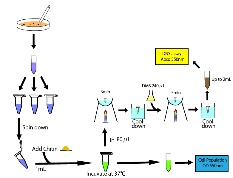
- added 240㎕ DNS reagent to 80㎕sample
- heated it for 5 min in boiling water and then cooled it in water.
- added it distilled water by 2ml
- measured absorbance in 550nm
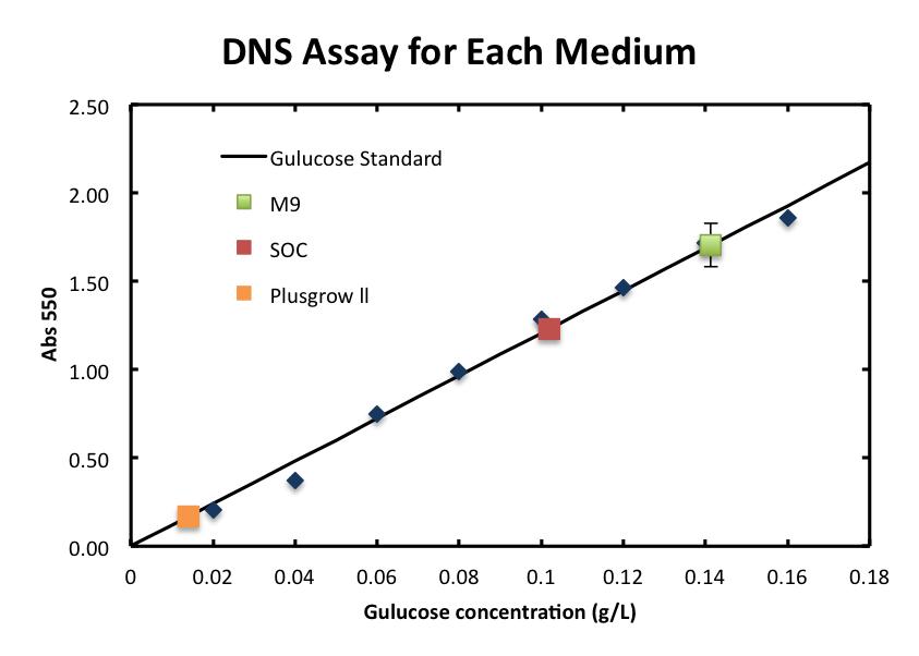
The standard curve is shown in fig 1.
1-2:evaluate the affection of medium
we examined the interruption based on the components of media(SOC, plas-grow and M9)
| Sample | each medium |
| Blank | mixed liquid (240μℓDNS reagent plus 1760㎕distilled water ) |
- added 240㎕ DNS reagent to 80㎕sample
- heated it for 5 min in boiling water and then cooled it in water.
- added it distilled water by 2ml
- measured absorbance in 550nm
The result is shown in fig 2.
1-3:measurement the effect of remainded E.coli
| Sample | each medium |
| Blank | mixed liquid (240μℓDNS reagent plus 1760㎕distilled water ) |
This assay was performed three times for each medium
- poured the medium cultured E.coli overnight 1.2 ㎕ into each five microcentritube.
- centrifuged them for 5 min at 5,000 rpm
- prepared new five microcentritube and move 800㎕ the supernatants into each of them.
- measure the OD550 of one tube(use fresh medium as a blank in following assays)
- one hour after, we measured OD 550 of other tube
- take 80㎕ supernatant and move it into new tube and then heated it for 3 min in boiling water and then cooled it in water.
- added 240㎕ DNS reagent and heated it for 3 min in boiling water and then cooled it in water.
- two, three, five hours after, we did above operation, taking supernatant, measured OD500, heating and cooling, applying DNS reagent and heating and cooling again.
- added all sample tube (containing 320㎕ solution) distilled water by 2ml and measure the absorbance of them in 550nm.
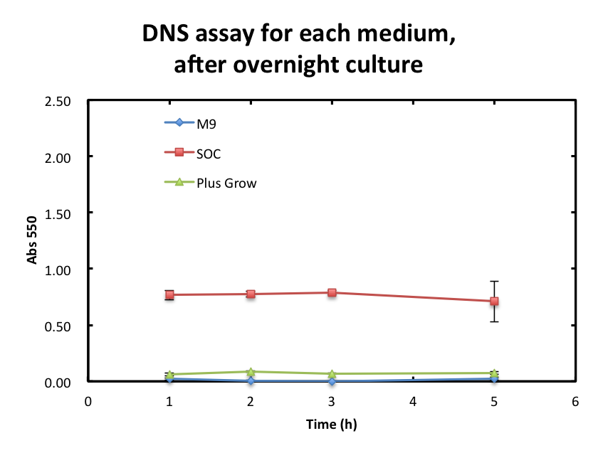
The results of measurement OD550 are shown in fig 3 and of measurement DNS assay are shown in fig 4.
Result
1. Standard Measurement for ChiA1.
- From the result, a strong correlation between glucose concentration and its A550 was observed.
2. Consideration of medium and growth of E.coli.
- We checked the influence of each medium to the DNS assay. Figure 2 shows the background absorbance of each medium. The absorbance of M9 was 1.7±0.1, SOC was1.227±0.007, and Plusgrow Ⅱ was 0.17±0.02.
Discussion
To overcome these barriers, we decided detail plan of our assay. From the result fig 3, SOC medium cultured E.coli overnight would still include too much amount of reducing materials and, from fig 3. plas-grow enabled reminded E.coli to grow rapidly. However, as for M9 medium, the increase of absorbance was lowest and there were fewest E.coli after 5 hour culture. So we choose M9 as the medium we will used in our assay. Another barrier, interruption of remained E.coli, can be avoided by the following way, conducting DNS assay before chitin is applied and after chitin is decomposed. The difference of these two assays’ data will show the real activity of chitinase.
Reference
[1] “Actinobacteria.” Internet: http://en.wikipedia.org/wiki/Actinobacteria [Nov. 5, 2011]
[2] H. Ikeda, J. Ishikawa, A. Hanamoto, M. Shinose, H. Kikuchi, T. Shiba, Y. Sakaki, M. Hattori, S. Omura, “Complete genome sequence and comparative analysis of the industrial microorganism Streptomyces avermitilis.” Nat Biotechnol., vol. 21, no. 5 pp. 526-31, Apr. 2003
[3] S. Omura, H. Ikeda, J. Ishikawa, A. Hanamoto, C. Takahashi, M. Shinose, Y. Takahashi, H. Horikawa, H. Nakazawa, T. Osonoe, H. Kikuchi, T. Shiba, Y. Sakaki, M. Hattori, “Genome sequence of an industrial microorganism Streptomyces avermitilis: deducing the ability of producing secondary metabolites.” Proc Natl Acad Sci U S A. vol. 98, no. 21 pp. 12215-20, Oct. 9
[4] H. Sakuzou, ”還元糖の定量法(生物化学実験法)” Kyoto University: Japan Scientific Soceties Press
[5] S. Kongruang, M. J. Han, C. I. Breton, M. H. Penner, “Quantitative Analysis of Cellulose-Reducing Ends.” Appl Biochem Biotechnol. Vol. 113, no. 116 pp. 213-31, Spring 2004
 "
"


