Team:UCL London/Research/Supercoiliology/Theory
From 2011.igem.org
The Theory of Gyrase
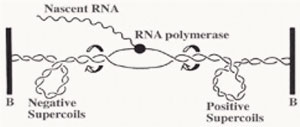
DNA gyrase is discovered in 1976 by a group of researchers (Martin Gellert et.al.) in the National Institutes of Health, Bethesda ,Marryland, USA . The prokaryotic gyrase is found to be the only type of type II topoisomerase which is able to introduce negative supercoils into the closed circular plasmid. The reactions requires magnesium ions and adenosine triphosphate (ATP) as the energy source. DNA gyrase plays an important role in the growth as well as the replication of the E.coli bacteria. A series of report show that DNA gyrase relaxes the stress which is build up downstream of the gene transcription and DNA replication . The positive supercoils stress which builds up is removed by DNA gyrases so that transcription or DNA replication can be allowed to proceed without having the opposing downstream stress to stop the RNA or DNA polymerase.
Negative supercoiled plasmid also allows easier transition from the closed promoter complex to the open promoter complex and this will allow higher transcription efficiency from the underwound B-DNA(right handed helix). Although in this theory , most promoters will have better transcriptional efficiency , but there are exceptions in this theory.
In E.coli, the level of supercoiling act as a regulatory mechanism . Gyrase genes will be repressed when the negative supercoiling level exceed a certain threshold and this observation is believed to be due to the conformation adopted by the DNA sequence flanking the gyrase genes and promoters .The transcription of gyrA and gyrB encoding the subunits of DNA gyrase is induced by relaxation of DNA. It has been proposed that promoter clearance but not open complex formation is the rate-limiting step for these promoters. When negative supercoiling achieve a very high level, the expression of DNA gyrase will be repressed while expression of topoisomerase I will be upregulated. Topoisomerase I will be responsible for the relaxation of the negative supercoiled DNA. Hence due to this regulatory mechanism the state of supercoiling in the E.coli cell will be in an equilibrium, According to a few research papers, the superhelical density (indicated by sigma symbol) of the E.coli chromosome is maintained at physiological level of ca. -0.05 as first measured by SInden et. al.(1980), by the combined action of gyrase, topo I and topoIV.
PICTURE GYRASE ACTION MECHANISM
It has been proposed that the tetrameric gyrase would bind to the gyrase binding site in the DNA to form a gyrase-DNA complex with about 120bp of DNA wrapped around the protein in a positive supercoiling sense. Since gyrase-DNA complex introduces a positive supercoil into the closed circular plasmid, there must be another negative supercoil (writhe) formed within the plasmid to balance out the positive supercoil as the linking number of the plasmid would remain constant unless DNA strand breakage and resealing occurred.
Next gyrA will break the DNA strand and the Tyr122 residue will form a phosphotyrosine linkage with the cleaved 5’ end of backbone while the 3’ end will be held by non-covalent forces between the enzyme and the DNA . Each gyrA subunits will form one phophotyrosine linkage with each strand and allow the DNA strand to pass through the breakage gap and after that the DNA strands resealed through a nucleophilic attack between the oxygen in the 3’ hydroxyl group and the phosphotyrosine linkage of the 5’ end. In this way gyrase reduces linking number by two in the plasmid. Thus, a single cycle of reaction occurred with two ATP consumed by two gyrB subunits.
In the cycle of reaction , divalent cations such as Mg2+ ions are essential to bind to the beta and gamma phosphate group of the ATP . This is to allow the correct binding of ATP substrate to the active site in gyrB subunits. Thus, by introducing suitable chelating agent into the reaction mixture , one will observe the inhibition of the gyrase activity . By using AMP-PNP which is an analogue of the ATP, gyrB subunits which bind to the molecule can be visualised by using the X-ray crystallography technique. An image which visualize the model is provided in the wiki.
Here, the main aim of the gyrase project is to extract and create a gyrase subunits expression biobrick composite part so that we can stimulate the overproduction of gyrase proteins in the transformed cells. By using EcoCyc and NCBI , we have identify the gyrase A and gyrase B genes which are responsible for the expression of the functional gyrase unit that is composed of two gyrA and two gyrB subunits. From here we have designed PCR primers with the standard restriction enzyme sites incorporated. EcoR1 and XbaI in the upstream part of the gyrase ORF while SpeI and PstI in the downstream part of the ORF.
</html>
 "
"

