Team:HKU-Hong Kong/Lab Diaries

From 2011.igem.org

Revision as of 17:12, 4 October 2011 by KarenSiu (Talk | contribs)

Lab Diaries
Week 1

Transformation of reporter DNA (pEGFP-loxp-km-loxp) into DH10B (non-virulent strain E. coli) with antibiotic resistance (Chloramphenicol – Cm)

Lab Diaries
Week 2
  1. tetO2 -1 (DNA binding site)
    1. Reverse PCR was used to insert tetO2 -1 into pEGFP-loxp-km-loxp (template DNA) by using forward and reverse primers which are with the tetO2 -1. By using reverse PCR, we can insert the tetO2 -1 site into the pEGFP while the plasmid produced is still in double-stranded circular form.
    2. pEGFP-loxp-km-loxp-tetO2-1 was then transformed into DH10B to greatly amplify the product by using bacterial cells
  2. tetO2 -2 (DNA biding site)
    1. Reverse PCR was used to insert tetO2 -2 into pEGFP-loxp-km-loxp
    2. 2uL of 10-fold diluted pEGFP-loxp-km-loxp- tetO2 -2 was transformed into DH10B to greatly amplify the product by using bacterial cells
  3. tetO2 – 3 (DNA binding site)
    1. Reverse PCR was used to insert tetO2 – 3 into pEGFP-loxp-km-loxp
    2. 2uL of 10-fold diluted pEGFP-loxp-km-loxp- tetO2 – 3 was transformed into DH10B to greatly amplify the product by using bacterial cells
Lab Diaries
Week 3
  1. Overlap PCR (involving 3 steps) was carried out to produce the fusion protein [tetR-HNS (any length)]
    1. PCR was carried out separately for 2 genes, tetR and HNS (full length, in this case)
      1. Primers used were as below:
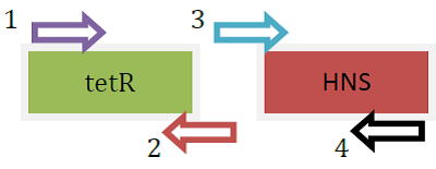
      2. tetR: [forward] R-H-out-F; [reverse] R-H-tetR-R
      3. HNS (FL): [forward] R-H-tetR-F-out; [reverse] R-H(FL)-2-R
        KAREN Fusion protein gene.png
        Fusion protein genes produced separately using PCR
    2. Another PCR was carried out using primers with linker to insert the linker DNA to the tetR and HNS (FL) for the fusion of the 2 genes.
    3. PCR was used to further amplify the product from step 2 (fusion protein gene)
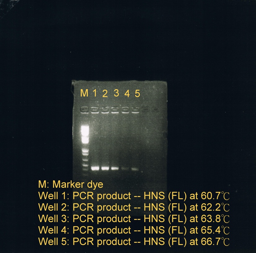
  2. PCR was used to obtain HNS(FL) at 5 different temperatures to test which temperature is optimal for annealing
    1. Annealing phase at 5 different temperatures: (a) 60.7C; (b) 62.2C; (c) 63.8C; (d) 65.4C; (e) 66.7C (less sample)
    2. Electrophoresis was carried to determine which temperature best suit the annealing phase. From the gel image, it was observed that a high concentration of products was resulted at annealing temperature between 60.7C and 62.2C.
      KAREN fusion ptn 2.jpg
      Fusion protein genes annealed at 5 different temperatures