Team:Virginia Tech
From 2011.igem.org
| Line 29: | Line 29: | ||
<h2>Characterization of Fluorescent Reporters:</h2> | <h2>Characterization of Fluorescent Reporters:</h2> | ||
| - | [[File:division.jpg|frame | + | [[File:division.jpg|frame 500 px|100px|right]] |
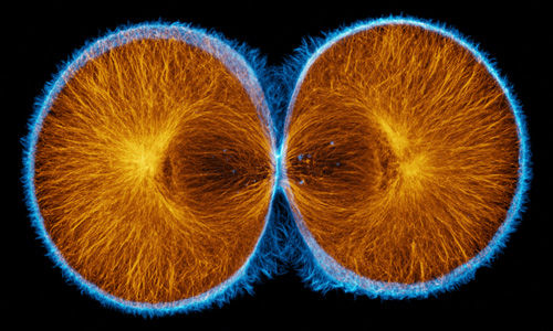
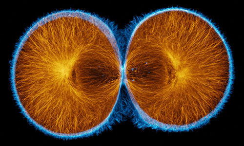
Fluorescent proteins have become ubiquitous tools for studying cellular processes, and are frequently used to visualize the dynamics of synthetic networks in cells. To be particularly effective for these applications, fluorescent proteins must feature fast maturation and degradation rates, and these rates must be well-characterized and documented. The 2011 VT iGEM team has worked to find and characterize fluorescent proteins and degradation tags that more quickly degrade them. Here, we present chemical and mathematical models based on two parameters, maturation and degradation rates, and in doing so, we explored difficulties in the process of characterizing parts. In conducting our experimentation, we tested fluorescent proteins with different degradation tags in <i>Escherichia coli </i> and <i>Saccharomyces cerevisiae</i> using automated fluorescent microscopy techniques, and then worked to determine a comprehensive, accurate mathematical basis for fluorescent protein characterization. | Fluorescent proteins have become ubiquitous tools for studying cellular processes, and are frequently used to visualize the dynamics of synthetic networks in cells. To be particularly effective for these applications, fluorescent proteins must feature fast maturation and degradation rates, and these rates must be well-characterized and documented. The 2011 VT iGEM team has worked to find and characterize fluorescent proteins and degradation tags that more quickly degrade them. Here, we present chemical and mathematical models based on two parameters, maturation and degradation rates, and in doing so, we explored difficulties in the process of characterizing parts. In conducting our experimentation, we tested fluorescent proteins with different degradation tags in <i>Escherichia coli </i> and <i>Saccharomyces cerevisiae</i> using automated fluorescent microscopy techniques, and then worked to determine a comprehensive, accurate mathematical basis for fluorescent protein characterization. | ||
Revision as of 01:36, 29 September 2011
| Home | Team | Official Team Profile | Project | Parts Submitted to the Registry | Notebook | Safety | Attributions |
|---|
 "
"


