Team:Lethbridge/Results

From 2011.igem.org

(Difference between revisions)
Line 33: Line 33:
<br>
<br>
<BLOCKQUOTE>
<BLOCKQUOTE>
 +
 +
=Part: BBa_K542010 - Enhanced Lumazine Synthase (ELS)=
 +
 +
Datasheet for <html><a href="http://partsregistry.org/Part:BBa_K542010"><font color="blue">Part BBa_I716462</font></a></html> in <i>E. coli</i> strain BL21 (DE3).
 +
<br>
 +
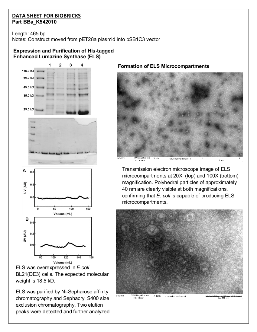
[[image:uoflELSdatasheet.png|center|150px]]
 +
 +
==Introduction==
 +
Lumazine synthase (LS) from <i>Aquifex aeolicus</i> forms icosahedral microcompartment (MC) assemblies of 60 or 180 monomeric units that self assemble and are capable of isolating proteins from their local environment. As shown in previously published work (1), the LS protein has been mutated so that the interior of the MC is negatively charged; the UL 2009 iGEM team has submitted the mutated LS gene to the parts registry (BBa_K249002). A negatively charged interior allows for preferential compartmentalization of positively charged molecules, which can easily be engineered through the addition of a poly-arginine tag to a target protein (1). The size of the cavity of the Lumazine Synthase microcompartment was enlarged and the negative charge was increased via directed evolution (2). The compartment formed by this polypeptide has a larger cavity and a larger net negative charge, thus allowing a larger loading capacity into the cavity of the compartment.
 +
<br><br>
 +
The part was synthesized by Bio Basic Inc. into the pET28a plasmid vector (which allows for expression of ELS with an N-terminal His-tag). The Enhanced Lumazine Synthase (ELS) was moved into the pSB1C3 vector by the Lethbridge 2011 team.
 +
==Characterization==
 +
===Overexpression===
 +
The His-tagged ELS in pET28a was transformed into <i>E. coli</i> BL21 (DE3) and overexpressed.
 +
====Materials and Methods====
 +
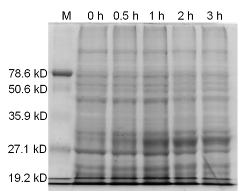
4 X 500 mL of LB media in 2 L flasks were inoculated with <i>E. coli</i> BL21 (DE3) cells expressing ELS. Cells were grown at 37°C with shaking until the culture reached an OD<sub>600</sub> of 0.683. The cultures were then induced with 10 mM IPTG. Culture samples containing equal amounts of cells were taken before induction and 30 min, 1 hour, 2 hours, and 3 hours post induction for analysis by SDS-PAGE. At 5 hours post induction the cells were harvested by centrifugation at 5000 x g for 5 min. The cell lysate was resuspended in 42 mL of buffer containing 50 mM Tris pH 8.0, 60 mM NH<sub>4</sub>CL, 7 mM β-mercaptoethanol, 1 mM PMSF, 7 mM MgCl<sub>2</sub>, 300 mM KCl, 10 mM imidazole, and 15% glycerol. Afterwards, 0.05 g of crystallized lysozyme was added to the cell suspension and incubated on ice for 1 hour. The lysate was then centrifuged for 70 minutes at 30000 x g to obtain the S30 fraction.
 +
====Results====
 +
[[image:uoflELSexpressiongel.png|center]]
 +
<br><br>
 +
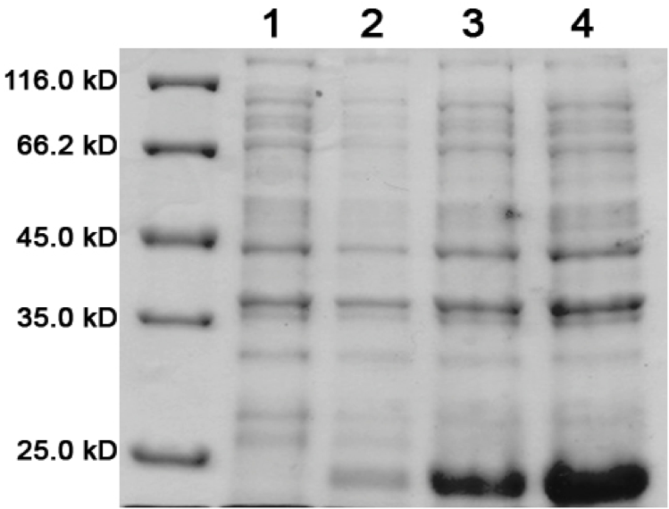
<b>Figure 1.</b>    12.5% SDS-PAGE of His-tagged ELS Overexpressed in <i>E. coli</i> BL21 (DE3). Samples were taken at 1 hour increments after IPTG induction. Lane 1: zero hours; Lane 2: 1 hour; Lane 3: 2 hours; Lane 4: 3 hours. The expected molecular weight of enhanced lumazine synthase is 18.5 kD.
 +
====Conclusion====
 +
Figure 1 clearly displays an increase in expression of a protein smaller than 25.0 kD, which is approximately the expected size of enhanced lumazine synthase monomers (18.5 kD).
 +
===Protein Purification===
 +
Enhanced lumazine synthase microcompartments were purified using size exclusion chromatography (SEC) to determine if the purified ELS formed a homogenous mixture of compartments, heterogenous mixture of microcompartments, or remained in monomer form.
 +
====Materials and Methods====
 +
In order to obtain a sample of ELS that was near homogeneity for characterization with SEC, the ELS sample was first purified by immobilized metal-ion affinity chromatography (IMAC). Ni<sup>2+</sup>-Sepharose beads supplied by Sigma Aldrich were pre-swelled in buffer containing 20% ethanol and 1mM NaCl. 3.75 mL of the Ni<sup>2+</sup>-Sepharose slurry was added to a 50 mL falcon tube. The beads were centrifuged at 500 x g for 2 min and the ethanol-containing buffer decanted. The beads were washed with 3 bed volumes of sterile water. The column was then washed in 6 bed volumes of Buffer A containing 50 mM Tris pH 8.0, 60 mM NH<sub>4</sub>CL, 7 mM β-mercaptoethanol, 1 mM PMSF, 7 mM MgCl<sub>2</sub>, 300 mM KCl, 10 mM imidazole, and 15% glycerol. S30 cell extract was applied to the column and mixed gently. The resin-S30 extract suspension was incubated on ice for 1 hour. The mixture was centrifuged at 500 x g for 2 min and the supernatant decanted. The supernatant was kept for further analysis by SDS-PAGE. The resin was washed 3 times with a full falcon tube volume (50 mL) of Buffer A. The mixture was centrifuged at 500 x g for 2 min and the supernatant decanted. The supernatant was kept for further analysis by SDS-PAGE. The column was washed 4 times with a full falcon tube volume (50 mL) of Buffer B (Buffer A plus 20 mM imidazole). The mixture was centrifuged at 500 x g for 2 minutes and the supernatant decanted. The supernatant was kept for further analysis by SDS-PAGE. The protein was eluted 9 times using 90% bed volume of Buffer E (Buffer A plus 250 mM imidazole). After each wash the sample was kept for analysis by SDS-PAGE.
 +
<br><br>
 +
Fractions 1 – 9 were pooled and concentrated using a Vivaspin Column with a 10 kDa molecular weight cut-off.
 +
<br><br>
 +
Concentrated protein samples from the Ni<sup>2+</sup>-Sepharose column were applied to a Sephacryl S400 size exclusion chromatography column at a flow rate of 0.4 mL/minute. 150 mL of SEC Buffer (50 mM sodium phosphate, 5 mM EDTA, 200 mM NaCl, pH 8.0, with 20% glycerol) was pumped through the column. The fractions eluted were collected and the absorbance was measured at 280 nm (see Fig 3 and 4).
 +
====Results====
 +
[[image:uoflELSsdspageimac.png|center]]
 +
<br>
 +
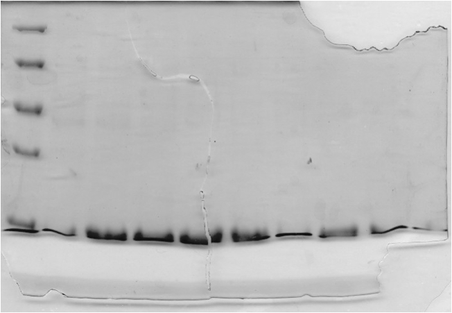
<b>Figure 2.</b>    SDS-PAGE (15%) of ELS purification by IMAC. Lane 1: protein marker. Lanes 2-10: Elution Fractions 1 – 9.
 +
<br><br>
 +
[[image:uoflELSchromatograms.png|center]]
 +
<br>
 +
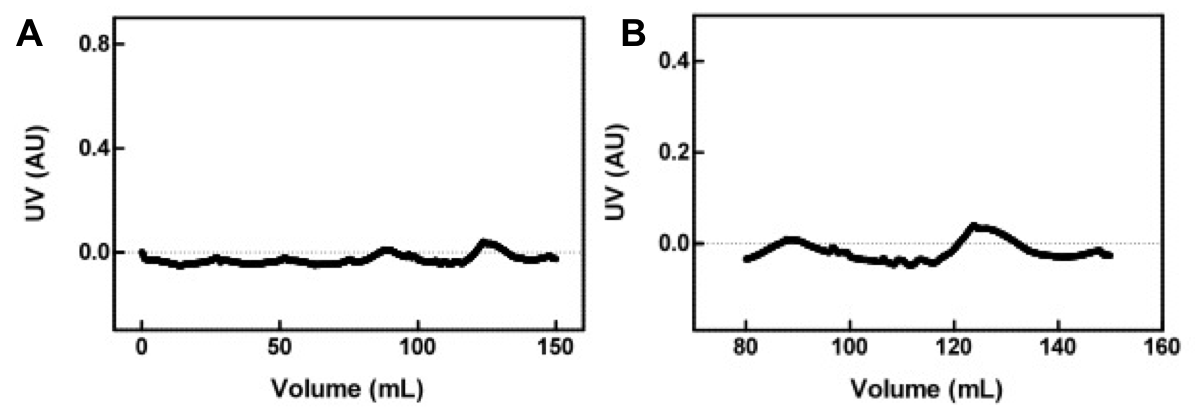
<b>Figure 3.</b>    (A) Concentrated protein sample from the Ni<sup>2+</sup>-Sepharose column applied to a Sephacryl S400 size exclusion chromatography column at a flow rate of 0.4 mL/minute. The absorbance of the eluting solution was measured at 280nm. (B) Magnified view of chromatogram of the two peaks seen in (A).
 +
====Conclusion====
 +
Figure 2 shows that with each wash proteins of a similar size were eluted off the resin. These proteins run at the expected molecular weight of enhanced lumazine synthase monomers (18.5 kD).
 +
<br><br>
 +
The chromatogram (Figure 3) from the chromatography experiment displays two regions of increased absorbance at 280 nm, between 82 and 135 mL of buffer eluted. These fractions were pooled, concentrated, and analyzed using transmission electron microscopy.
 +
===Transmission Electron Microscopy===
 +
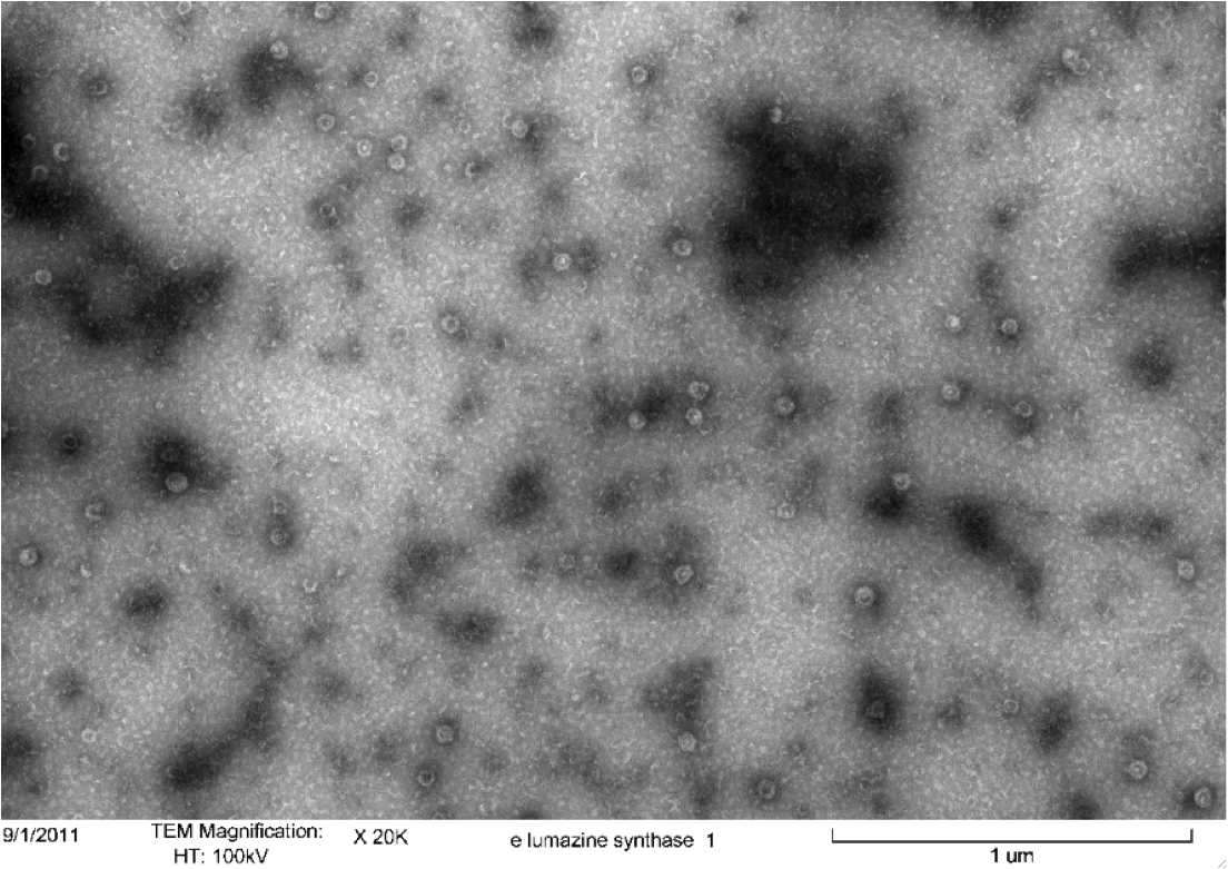
Purified samples of enhanced lumazine synthase from size exclusion chromatography (SEC) were characterized using transmission electron microscopy. We viewed samples of each of the two peaks from the SEC; however only one sample contained microcompartments.
 +
====Materials and Methods====
 +
Purified Samples in solution were placed on a carbon grid and negatively stained using uranyl acetate. Carbon grids containing the samples were then viewed with a Hitachi H-7500 Transmission Electron Microscope. 
 +
====Results====
 +
[[image:uoflELSTEM1.png|center]]
 +
<br>
 +
<b>Figure 4.</b>    Transmission electron microscopy of Enhanced Lumazine Synthase microcompartments.  Microcompartments of approximately 40 nm can be seen at x 20K magnification.
 +
<br><br>
 +
[[image:uoflELSchromatograms.png|center]]
 +
<br>
 +
<b>Figure 5.</b>    Transmission electron microscopy of Enhanced Lumazine Synthase microcompartments.  Microcompartments of approximately 40 nm can be seen at x 100K magnification.
 +
====Conclusion====
 +
As expected, TEM micrographs show polyhedral particles of approximately 40 nm, corresponding to the size described by Wörsdörfer et al. (2011). This confirms that E. coli is capable of producing enhanced lumazine synthase.
 +
==References==
 +
 +
(1) Viadiu H, Aggarwal AK (2000). Structure of BamHI bound to nonspecific DNA: a model for DNA sliding. Mol. Cell 5 (5): 889-895.
 +
<br>
 +
(2) iGEM 2007. (2011, May 10). BerkiGEM2007Present5. Retrieved from http://parts.mit.edu/igem07/index.php/BerkiGEM2007Present5.
 +
=<font color="black">Part: BBa_I716462 – <i>Bam</i>=

Revision as of 15:57, 28 September 2011


Contents

Part: BBa_K542010 - Enhanced Lumazine Synthase (ELS)

Datasheet for Part BBa_I716462 in E. coli strain BL21 (DE3).

UoflELSdatasheet.png

Introduction

Lumazine synthase (LS) from Aquifex aeolicus forms icosahedral microcompartment (MC) assemblies of 60 or 180 monomeric units that self assemble and are capable of isolating proteins from their local environment. As shown in previously published work (1), the LS protein has been mutated so that the interior of the MC is negatively charged; the UL 2009 iGEM team has submitted the mutated LS gene to the parts registry (BBa_K249002). A negatively charged interior allows for preferential compartmentalization of positively charged molecules, which can easily be engineered through the addition of a poly-arginine tag to a target protein (1). The size of the cavity of the Lumazine Synthase microcompartment was enlarged and the negative charge was increased via directed evolution (2). The compartment formed by this polypeptide has a larger cavity and a larger net negative charge, thus allowing a larger loading capacity into the cavity of the compartment.

The part was synthesized by Bio Basic Inc. into the pET28a plasmid vector (which allows for expression of ELS with an N-terminal His-tag). The Enhanced Lumazine Synthase (ELS) was moved into the pSB1C3 vector by the Lethbridge 2011 team.

Characterization

Overexpression

The His-tagged ELS in pET28a was transformed into E. coli BL21 (DE3) and overexpressed.

Materials and Methods

4 X 500 mL of LB media in 2 L flasks were inoculated with E. coli BL21 (DE3) cells expressing ELS. Cells were grown at 37°C with shaking until the culture reached an OD600 of 0.683. The cultures were then induced with 10 mM IPTG. Culture samples containing equal amounts of cells were taken before induction and 30 min, 1 hour, 2 hours, and 3 hours post induction for analysis by SDS-PAGE. At 5 hours post induction the cells were harvested by centrifugation at 5000 x g for 5 min. The cell lysate was resuspended in 42 mL of buffer containing 50 mM Tris pH 8.0, 60 mM NH4CL, 7 mM β-mercaptoethanol, 1 mM PMSF, 7 mM MgCl2, 300 mM KCl, 10 mM imidazole, and 15% glycerol. Afterwards, 0.05 g of crystallized lysozyme was added to the cell suspension and incubated on ice for 1 hour. The lysate was then centrifuged for 70 minutes at 30000 x g to obtain the S30 fraction.

Results

UoflELSexpressiongel.png


Figure 1. 12.5% SDS-PAGE of His-tagged ELS Overexpressed in E. coli BL21 (DE3). Samples were taken at 1 hour increments after IPTG induction. Lane 1: zero hours; Lane 2: 1 hour; Lane 3: 2 hours; Lane 4: 3 hours. The expected molecular weight of enhanced lumazine synthase is 18.5 kD.

Conclusion

Figure 1 clearly displays an increase in expression of a protein smaller than 25.0 kD, which is approximately the expected size of enhanced lumazine synthase monomers (18.5 kD).

Protein Purification

Enhanced lumazine synthase microcompartments were purified using size exclusion chromatography (SEC) to determine if the purified ELS formed a homogenous mixture of compartments, heterogenous mixture of microcompartments, or remained in monomer form.

Materials and Methods

In order to obtain a sample of ELS that was near homogeneity for characterization with SEC, the ELS sample was first purified by immobilized metal-ion affinity chromatography (IMAC). Ni2+-Sepharose beads supplied by Sigma Aldrich were pre-swelled in buffer containing 20% ethanol and 1mM NaCl. 3.75 mL of the Ni2+-Sepharose slurry was added to a 50 mL falcon tube. The beads were centrifuged at 500 x g for 2 min and the ethanol-containing buffer decanted. The beads were washed with 3 bed volumes of sterile water. The column was then washed in 6 bed volumes of Buffer A containing 50 mM Tris pH 8.0, 60 mM NH4CL, 7 mM β-mercaptoethanol, 1 mM PMSF, 7 mM MgCl2, 300 mM KCl, 10 mM imidazole, and 15% glycerol. S30 cell extract was applied to the column and mixed gently. The resin-S30 extract suspension was incubated on ice for 1 hour. The mixture was centrifuged at 500 x g for 2 min and the supernatant decanted. The supernatant was kept for further analysis by SDS-PAGE. The resin was washed 3 times with a full falcon tube volume (50 mL) of Buffer A. The mixture was centrifuged at 500 x g for 2 min and the supernatant decanted. The supernatant was kept for further analysis by SDS-PAGE. The column was washed 4 times with a full falcon tube volume (50 mL) of Buffer B (Buffer A plus 20 mM imidazole). The mixture was centrifuged at 500 x g for 2 minutes and the supernatant decanted. The supernatant was kept for further analysis by SDS-PAGE. The protein was eluted 9 times using 90% bed volume of Buffer E (Buffer A plus 250 mM imidazole). After each wash the sample was kept for analysis by SDS-PAGE.

Fractions 1 – 9 were pooled and concentrated using a Vivaspin Column with a 10 kDa molecular weight cut-off.

Concentrated protein samples from the Ni2+-Sepharose column were applied to a Sephacryl S400 size exclusion chromatography column at a flow rate of 0.4 mL/minute. 150 mL of SEC Buffer (50 mM sodium phosphate, 5 mM EDTA, 200 mM NaCl, pH 8.0, with 20% glycerol) was pumped through the column. The fractions eluted were collected and the absorbance was measured at 280 nm (see Fig 3 and 4).

Results

UoflELSsdspageimac.png


Figure 2. SDS-PAGE (15%) of ELS purification by IMAC. Lane 1: protein marker. Lanes 2-10: Elution Fractions 1 – 9.

UoflELSchromatograms.png


Figure 3. (A) Concentrated protein sample from the Ni2+-Sepharose column applied to a Sephacryl S400 size exclusion chromatography column at a flow rate of 0.4 mL/minute. The absorbance of the eluting solution was measured at 280nm. (B) Magnified view of chromatogram of the two peaks seen in (A).

Conclusion

Figure 2 shows that with each wash proteins of a similar size were eluted off the resin. These proteins run at the expected molecular weight of enhanced lumazine synthase monomers (18.5 kD).

The chromatogram (Figure 3) from the chromatography experiment displays two regions of increased absorbance at 280 nm, between 82 and 135 mL of buffer eluted. These fractions were pooled, concentrated, and analyzed using transmission electron microscopy.

Transmission Electron Microscopy

Purified samples of enhanced lumazine synthase from size exclusion chromatography (SEC) were characterized using transmission electron microscopy. We viewed samples of each of the two peaks from the SEC; however only one sample contained microcompartments.

Materials and Methods

Purified Samples in solution were placed on a carbon grid and negatively stained using uranyl acetate. Carbon grids containing the samples were then viewed with a Hitachi H-7500 Transmission Electron Microscope. 

Results

UoflELSTEM1.png


Figure 4. Transmission electron microscopy of Enhanced Lumazine Synthase microcompartments.  Microcompartments of approximately 40 nm can be seen at x 20K magnification.

UoflELSchromatograms.png


Figure 5. Transmission electron microscopy of Enhanced Lumazine Synthase microcompartments.  Microcompartments of approximately 40 nm can be seen at x 100K magnification.

Conclusion

As expected, TEM micrographs show polyhedral particles of approximately 40 nm, corresponding to the size described by Wörsdörfer et al. (2011). This confirms that E. coli is capable of producing enhanced lumazine synthase.

References

(1) Viadiu H, Aggarwal AK (2000). Structure of BamHI bound to nonspecific DNA: a model for DNA sliding. Mol. Cell 5 (5): 889-895.
(2) iGEM 2007. (2011, May 10). BerkiGEM2007Present5. Retrieved from http://parts.mit.edu/igem07/index.php/BerkiGEM2007Present5.


Part: BBa_I716462 – Bam

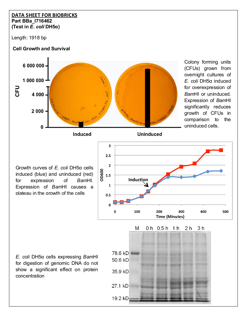
Datasheet for Part BBa_I716462 in E. coli strain DH5alpha.

UoflBamHIdatasheet.png

Introduction

Part I716462 was previously characterized by the UC Berkeley iGEM team in 2007. The purpose of this part was to prevent unwanted cell proliferation by engineering a genetic self-destruct mechanism into bacteria that upon induction would express a genetic material-degrading toxin, the endonuclease BamHI that would kill cells yet preserve their physical integrity. BamHI has a restriction cut site of G'GATCC, generating sticky ends (1). The restriction endonuclease was placed under the control of the arabinose- inducible promoter, pBAD. In 2007, the UC Berkeley iGEM team was able to show that cells lost their ability to reproduce but did not lyse upon induction with arabinose (2). The University of Lethbridge 2011 iGEM team wished to further characterize this part to degrade the genomic DNA of E. coli in order to have a functional chassis for our tailings pond clean-up kit without the risk of DNA contamination. In this way, tight control and regulation over our engineered bacterial cells is possible.

Characterization

Cell Survival

Materials and Methods

The BamHI construct was overexpressed in E. coli DH5α cells. Cultures were induced with arabinose at an OD600 of 0.6 and grown overnight. Samples of the induced culture and an uninduced culture were plated on LB agar plates to monitor growth of colony forming units.

Results

UoflBamHIgrowthcurve.png


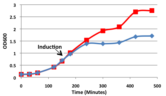
Figure 1. Growth curves of E. coli DH5α cells transformed with BamHI construct. Blue line represents cells induced at an OD600 of 0.60. Red line represents uninduced cells.

UoflBamHICFU.png


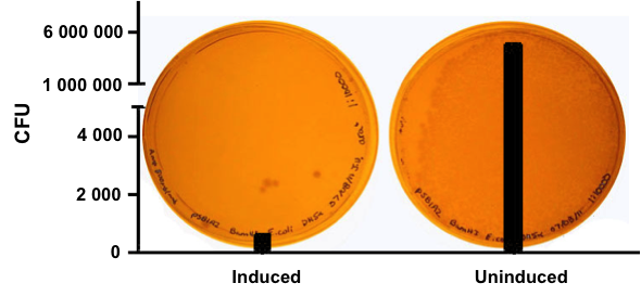
Figure 2. E.coliDH5α cells containing the BamHI construct from overnight cultures. The samples were plated on LB agar plates and the number of colony forming units (CFU) was counted. Left: culture induced for BamHI expression with arabinose, right: uninduced culture.

Analysis of Genomic DNA

Materials and Methods

E. coli DH5α cells were grown and induced to overexpress one of three samples: BamHI construct induced with arabinose (10 µM) at an OD600 of 0.6, BamHI construct uninduced, non-BamHI construct induced with arabinose (10 µM) at OD600 of 0.6. Cells were grown in LB media with 100 ng/mL ampicillin.

Qiagen Blood and Tissue Kit was used to isolate the genomic DNA of gram-negative bacteria. A 0.8% agarose gel was ran at 150 V for 90 minutes to separate the genomic DNA of E. coli DH5α cells collected at different time points (pre-induced and 30 min, 1 hr, 2 hr, and 3 hr post induction) for all three samples.

Results

UoflBamHIgenomicDNA.png


Figure 3. Isolation of genomic DNA from E. coli DH5α cells. From left to right: 1Kb + ladder, 3hr, 2 hr, 1 hr, and 30 min after induction with arabinose and pre-induction for cells containing BamHI gene construct, uninduced control, and cells not containing the BamH1 gene construct.

Conclusion

Upon induction with arabinose, E. coli DH5α cells containing the BamHI construct did not reproduce as the overexpression of the BamHI endonuclease digested the genomic DNA of the host cell. This is why in Figure 2 there are only 4 colonies seen as opposed to the control which has an uncountable amount of colonies. In Figure 1, the optical density of the BamHI expressing cells reaches a plateau; however, the same sample which has not been induced kept increasing, which indicates that the expression of BamHI is affecting normal growth of the cells.
Lastly, the genomic DNA of the E.coli cells was isolated. Upon overexpression of BamHI, there are still large segments of DNA at the pre-induced time point; however, as the post-induction time increases, there is an increase in digestion fragments.

References

(1) Viadiu H, Aggarwal AK (2000). Structure of BamHI bound to nonspecific DNA: a model for DNA sliding. Mol. Cell 5 (5): 889-895.
(2) iGEM 2007. (2011, May 10). BerkiGEM2007Present5. Retrieved from http://parts.mit.edu/igem07/index.php/BerkiGEM2007Present5.