Team:Freiburg/Results
From 2011.igem.org
(Difference between revisions)
(→commons) |
Juliimapril (Talk | contribs) (→Experimental setup) |
||
| (22 intermediate revisions not shown) | |||
| Line 2: | Line 2: | ||
=results= | =results= | ||
==<span style="color:grey;">Precipitator</span>== | ==<span style="color:grey;">Precipitator</span>== | ||
| - | + | ||
| - | + | ||
[http://partsregistry.org/Part:BBa_K608406 BBa_K608406] | [http://partsregistry.org/Part:BBa_K608406 BBa_K608406] | ||
| Line 77: | Line 76: | ||
| - | '''Precipitator fused with GST tag''' | + | |
| + | ===[https://2011.igem.org/Team:Freiburg/Modelling#A_mathematical_model_to_determine_the_experimental_design Mathematical modeling]=== | ||
| + | |||
| + | To determine the Affinity k_D, experiments to find out the binding affinity of the plastic binding domain are necessary. To get a direct access to these values, we cloned the plastic binding domain in front of a GFP. Then, dilution and washing assays could be performed on polystyrene microtiter plates, red out by a fluorescence plate reader. | ||
| + | The desired parameters could be calculated by measuring dilution rows of GFP proteins and measuring the fluorescence signals at the different concentrations. C_total could be determined by a dilution row with subsequent washing steps, to find out at what [P] concentration there is a saturation. See description of the plastic binding subproject for more detailed explanation on the experimental setup. | ||
| + | |||
| + | A qualitative experiment to prove that Nickel is binding the Precipitator is sufficient, since k_2 >> 1 and does not play a significant role in our setup. This experiment could have been done using a nanofilter that blocks protein but let through ions. The Nickel concentration of the flow through can then be measured. | ||
| + | |||
| + | Alternatively purification of the Precipitator by fusing it with a GST-tag could be done, to subsequently measure the absorbance of the protein, before and after adding Nickel to the solution. After Jordan 1974 a detectable change in the absorbance should be detectable after the complex is formed. A similar effect – a colorshift from white to blue - is visible when one prepares a Ni-NTA column. For this purpose we cloned the GST domain in front of the Precipitator. However towards the end of the project there was no more time to perfom these experiments. | ||
| + | |||
| + | ==References== | ||
| + | |||
| + | |||
| + | Tiandi Wei et.al.; "LRRML: a conformational database and an XML description of | ||
| + | leucine-rich repeats (LRRs)"; BMC Structural Biology 2008 doi: 10.1186/1472-6807-8-47 | ||
| + | |||
| + | |||
| + | Schmidt, Marc et.al.; "Crucial role for human Toll-like receptor 4 in the development of contact allergy to nickel"; | ||
| + | Nature 2010 | ||
| + | |||
| + | |||
| + | Letter, J.E. et.al.; "Complexing of Nickel( 11) by Cysteine, Tyrosine, | ||
| + | and Related Ligands and Evidence for Zwitterion Reactivity"; | ||
| + | Contribution from the Department of Chemistry, University of Alberta, | ||
| + | Edmonton, Alberta T6G 2G2, Canada. | ||
| + | |||
| + | |||
| + | Kajava, A.V.;"Structural Diversity of Leucine-rich Repeat Proteins" | ||
| + | J. Mol. Biol. (1998) 277, 519±527 | ||
| + | |||
| + | |||
| + | Kim, Ho Min et. al.; "Crystal Structure of the TLR4-MD-2 Complex with Bound Endotoxin Antagonist Eritoran" | ||
| + | DOI 10.1016/j.cell.2007.08.002 | ||
| + | |||
| + | ==='''Precipitator fused with GST tag'''=== | ||
The Precipitator was fused with the C-terminus of our GST tag in order to extract and further test the construct for its Nickel binding affinity. We successfully cloned it together using the Gibson Assembly and then subsequently pasted it into the iGEM vector. The submitted sequence was partially confirmed by sequencing. | The Precipitator was fused with the C-terminus of our GST tag in order to extract and further test the construct for its Nickel binding affinity. We successfully cloned it together using the Gibson Assembly and then subsequently pasted it into the iGEM vector. The submitted sequence was partially confirmed by sequencing. | ||
| Line 204: | Line 237: | ||
Sandra Harper, David W. Speicher (2008); „Expression and Purification of GST Fusion Proteins“; John Wiley and Sons, Inc.; DOI: 10.1002/0471140864.ps0606s52 | Sandra Harper, David W. Speicher (2008); „Expression and Purification of GST Fusion Proteins“; John Wiley and Sons, Inc.; DOI: 10.1002/0471140864.ps0606s52 | ||
| + | |||
===<span style="color:grey;">Plastic binding domain</span>=== | ===<span style="color:grey;">Plastic binding domain</span>=== | ||
| + | [http://partsregistry.org/Part:BBa_K608404 BBa_K608404] | ||
| + | IPTG-inducible Promoter with plastic binding domain-tagged GFP | ||
| + | |||
| + | ====Experimental setup==== | ||
| + | According to [https://2011.igem.org/Team:Freiburg/Description#Plastic_binding_domain Adey et al.] the plastic binding domain (pbd) binds to the polystyrene surface of micro titer plates (96 well plates). To investigate the binding properties of the plastic binding tag we started several spectroscopic assays using a plate reader (FLUOstar Omega) and polystyrene plates (Greiner bio one) To detect the fluorescence of the GFP tagged to the plastic binding domain we used black plates and a well scanning program measuring 10x10 spots in each well of the micro titer plate. We performed several washing steps to find out how much of the proteins can be found in the eluate and how much remains bound on the plate’s surface. To compare the pbd-tagged GFP to a normal GFP without special plastic binding ability we also measured GFP obtained via expression with our diverse PR (Promoter-Ribosome-binding-site constructs). To affirm the results obtained by fluorescence spectroscopy used the Bradford assay and screened for different protein concentrations in transparent polystyrene plates. | ||
| + | The calibration lines necessary for calculation of protein concentration can be found [[Media:Bradford+Fluorescence calibration.pdf|here]]. | ||
| + | ====Results==== | ||
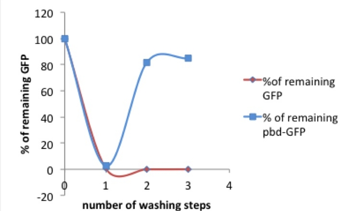
[[Image:picture 1 pbd.jpg|250px|right|thumb|Picture 1: % of tagged (red) or untagged (blue) GFP remaining in the well after washing compared to previous washing step / original concentration]] After the first measurement of the basic fluorescence intensity we transferred the samples onto another well, refilled the exhausted well with PBS and measured both eluate and remaining protein. In a second washing step the liquid was taken out of the first well again and given to another well. This washing was performed a third time, resulting in three eluates and a triply washed well with more or less protein remaining on the walls. | [[Image:picture 1 pbd.jpg|250px|right|thumb|Picture 1: % of tagged (red) or untagged (blue) GFP remaining in the well after washing compared to previous washing step / original concentration]] After the first measurement of the basic fluorescence intensity we transferred the samples onto another well, refilled the exhausted well with PBS and measured both eluate and remaining protein. In a second washing step the liquid was taken out of the first well again and given to another well. This washing was performed a third time, resulting in three eluates and a triply washed well with more or less protein remaining on the walls. | ||
<br/> | <br/> | ||
| Line 218: | Line 259: | ||
<br/> | <br/> | ||
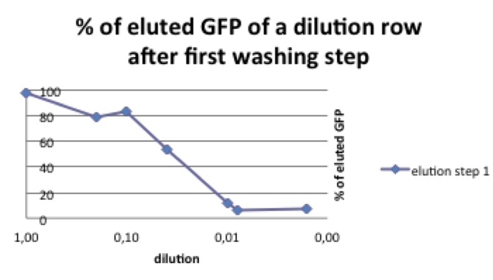
To investigate this phenomenon we compared different start concentrations of pbd-GFP concerning the amount of pbd-GFP that could be washed away. | To investigate this phenomenon we compared different start concentrations of pbd-GFP concerning the amount of pbd-GFP that could be washed away. | ||
| - | polystyrene micro titer plates provide only a limited surface for the pbd to bind, the solution shouldn’t be oversaturated with plastic binding protein. | + | polystyrene micro titer plates provide only a limited surface for the pbd to bind, the solution shouldn’t be oversaturated with plastic binding protein. In our case the diluted protein could only reach a surface of 91.5 mm2 per well. With a remaining protein amount of ~220 pg/well after three washing steps we estimate that 2,4 pg pbd-tagged protein can be bound per mm2. |
<br/> | <br/> | ||
| - | In a range of 1-30ng/µL start concentration there remains about seven times more pbd-GFP after washing than "normal" GFP (see picture 3). | + | In a range of 1-30ng/µL start concentration there remains about seven times more pbd-GFP after washing than "normal" GFP (see picture 3). |
| - | |||
| - | |||
<br/> | <br/> | ||
====Discussion==== | ====Discussion==== | ||
| - | + | In the experiments described above we could show that in a certain range of start concentration the plastic binding domain (pbd)-coupled GFP was more resistant to elution steps than GFP alone. This result indicates that the pbd-tagged GFP bound stronger to the plastic surface of the microtiter plate than “normal” GFP. The fact that in case of a rather high start concentration the majority of the pbd-GFP was washed away can be explained by the limitation of available binding surface. It seems like the pbd-GFP binds seven times better to the used polystyrene material than GFP alone. The amount of pbd-GFP that can bind to 1 mm2 is about 2,4 pg. To prove this supposition further measurement and more washing steps should be performed. | |
| Line 307: | Line 346: | ||
==<span style="color:grey;">Precipitator</span>== | ==<span style="color:grey;">Precipitator</span>== | ||
[http://partsregistry.org/Part:BBa_K608404 BBa_K608404] | [http://partsregistry.org/Part:BBa_K608404 BBa_K608404] | ||
| - | IPTG-inducible Promoter with plastic binding domain-tagged GFP | + | IPTG-inducible Promoter with '''plastic binding domain'''-tagged GFP |
[http://partsregistry.org/Part:BBa_K608406 BBa_K608406] | [http://partsregistry.org/Part:BBa_K608406 BBa_K608406] | ||
| Line 378: | Line 417: | ||
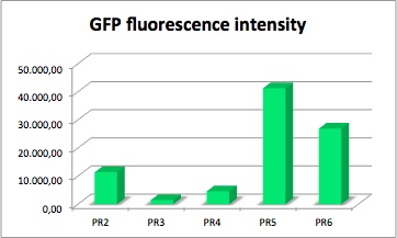
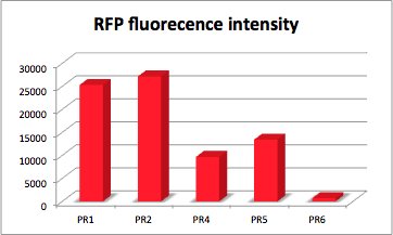
| - | Measurement of fluorescence intensity of PR-GFP and PR-RFP | + | '''Measurement of fluorescence intensity of PR-GFP and PR-RFP''' |
---- | ---- | ||
| Line 390: | Line 429: | ||
Samples were pipetted into the microplate and analyzed via the plate reader. In this experiment we focused on the protein concentration and the fluorescence intensity of RFP. | Samples were pipetted into the microplate and analyzed via the plate reader. In this experiment we focused on the protein concentration and the fluorescence intensity of RFP. | ||
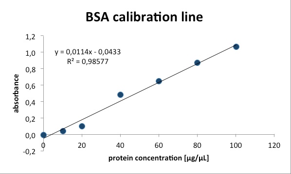
We measured the protein concentration with the bradford-assay. This is a method to determine the total protein concentration. To analyze the protein concentration of the samples, Coomassie Brillant Blue was pippeted to each sample. With the binding of the dye to the proteins the color changes from dark red to blue. The more protein in the solution the more Coomassie dye can bind to proteins and the more the color changes into blue. The absorption of bound Coomassie dye is 595nm. The absorbance is proportional with the amount of bound dye. With a series of Bovine Serum Albumin (BSA) measurements the exact protein concentration of the samples can be determined. BSA acts like a “marker” because the concentration of BSA is known and with a linear calibration line the exact protein concentration can be detected. | We measured the protein concentration with the bradford-assay. This is a method to determine the total protein concentration. To analyze the protein concentration of the samples, Coomassie Brillant Blue was pippeted to each sample. With the binding of the dye to the proteins the color changes from dark red to blue. The more protein in the solution the more Coomassie dye can bind to proteins and the more the color changes into blue. The absorption of bound Coomassie dye is 595nm. The absorbance is proportional with the amount of bound dye. With a series of Bovine Serum Albumin (BSA) measurements the exact protein concentration of the samples can be determined. BSA acts like a “marker” because the concentration of BSA is known and with a linear calibration line the exact protein concentration can be detected. | ||
| + | |||
| + | The values of the fluorescence intensity of RFP and GFP deviate from the expected values. We´ve expected PR1 to have the strongest GFP/RFP signal and PR6 with the lowest GFP/RFP signal. The data shows the differences. We could not repeat this measurement another time because of lack of time. We could not measure PR1-GFP because the sequencing was negative and there was no time for repeating. The measurement of PR3-RFP was an error and was excluded. | ||
'''PR-GFP''' | '''PR-GFP''' | ||
| Line 398: | Line 439: | ||
GFP served as a reporter of expression. We wanted to know how strong the promoter and RBS activity is. With this reporter gene it was possible to analyze the expression via plate reader. GFP is excited at a wavelength of 509nm and has an emission of 520nm. The plate reader illuminates the samples with a high energy xenon flash lamp. Optical filters or monochromator create the exact wavelength. The more GFP in the sample the higher is the GFP fluorescence intensity. The intensity is collected with the second optical system and is detected with a side window photomultiplier tube. | GFP served as a reporter of expression. We wanted to know how strong the promoter and RBS activity is. With this reporter gene it was possible to analyze the expression via plate reader. GFP is excited at a wavelength of 509nm and has an emission of 520nm. The plate reader illuminates the samples with a high energy xenon flash lamp. Optical filters or monochromator create the exact wavelength. The more GFP in the sample the higher is the GFP fluorescence intensity. The intensity is collected with the second optical system and is detected with a side window photomultiplier tube. | ||
| - | + | '''parts:'''<br/> | |
[http://partsregistry.org/Part:BBa_K608008 BBa_K608008] | [http://partsregistry.org/Part:BBa_K608008 BBa_K608008] | ||
constitutive strong promoter with medium RBS and GFP (PR2-GFP) | constitutive strong promoter with medium RBS and GFP (PR2-GFP) | ||
| Line 424: | Line 465: | ||
| - | + | '''parts:'''<br/> | |
[http://partsregistry.org/Part:BBa_K608013 BBa_K608013] | [http://partsregistry.org/Part:BBa_K608013 BBa_K608013] | ||
Latest revision as of 03:54, 22 September 2011
 "
"
 Contact


