Team:ETH Zurich/Process/Microfluidics

From 2011.igem.org

Revision as of 01:29, 22 September 2011 by Luwidmer (Talk | contribs)

Menu image preload Menu image preload Menu image preload Menu image preload Menu image preload Menu image preload


Microfluidics
First Channel Designs Final Channel Design

We relatively early figured out that we need a some kind of channel to establish the acetaldehyde or xylene gradient needed for SmoColi (see Circuit Design). However, there were several different channel designs possible, and the final design evolved through an iterative series of design steps and design validations. The first designs were validated based on vast simulations, the final design furthermore by biological experiments in the lab (see Systems Validation).

First Channel Designs

Microfluidic channel with flow and recycling of the medium

We came up with two different possible microfluidic channel designs, both involving a flow of toxic molecule in medium through the channel:

  • Variant 1: Plate with pixel filled with agarose and cell and a microfluidic channel above. This channel design consists of two parts: A PDMS plate with cubic "pixels" that is filled with cells in agarose by letting the liquid cell-agarose solution flow over the pixels by gravity. The agarose is then cooled down and the solidified surplus cell-agarose gel scraped off. A larger channel that is several pixels wide is then laid on top of the
Experimental setup for SmoColi.
Evaluation of the model.
  • Variant 2: Microfluidic channel with cells sitting in pockets in the channel.
Experimental setup for SmoColi.

Final Channel Design

Microfluidic channel without flow

Experimental setup for SmoColi.
Modeling showed that diffusion and degradation of acetaldehyde/ xylene is enough to create a concentration gradient in the tube. Without a flow there is no need for a liquid so we decided to fill the whole channel with agarose and cells likewise we don´t need recycling because AHL can diffuse through the whole channel.
Photo of the channel in action. The channel (the long thin tube at the right) is physically attached to a reservoir filled with the sample medium containing acetaldehyde or xylene (the Eppendorf tube at the lower left). In the case of acetaldehyde, the whole setup is packed in an impermeable plastic bag to significantly reduce the vaporization of acetaldehyde (not shown).
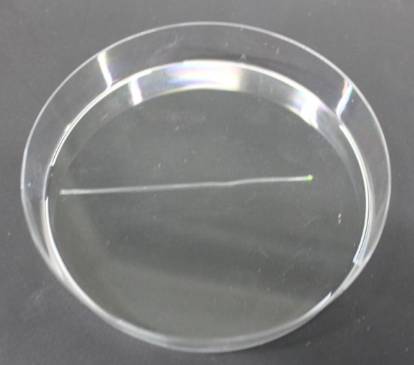
After the experiment, the physical channel is removed from the agerose medium containing the cells. The bald interior of the channel is placed on a petri dish (see picture) and analyzed under a fluorescence microscope.