Team:Cambridge/Future/Visuals
From 2011.igem.org
(Difference between revisions)
(Created page with "{{Template:Team:Cambridge/CAM_2011_TEMPLATE_HEAD}} As this project is based around colour and light interactions, our characterisation experiments are set up to provide visual r...") |
|||
| Line 4: | Line 4: | ||
==In vivo imaging== | ==In vivo imaging== | ||
| + | |||
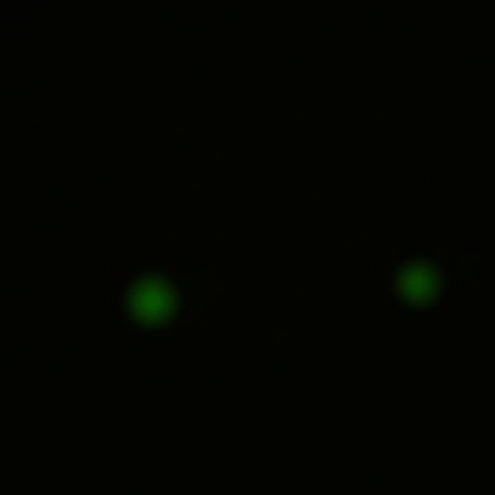
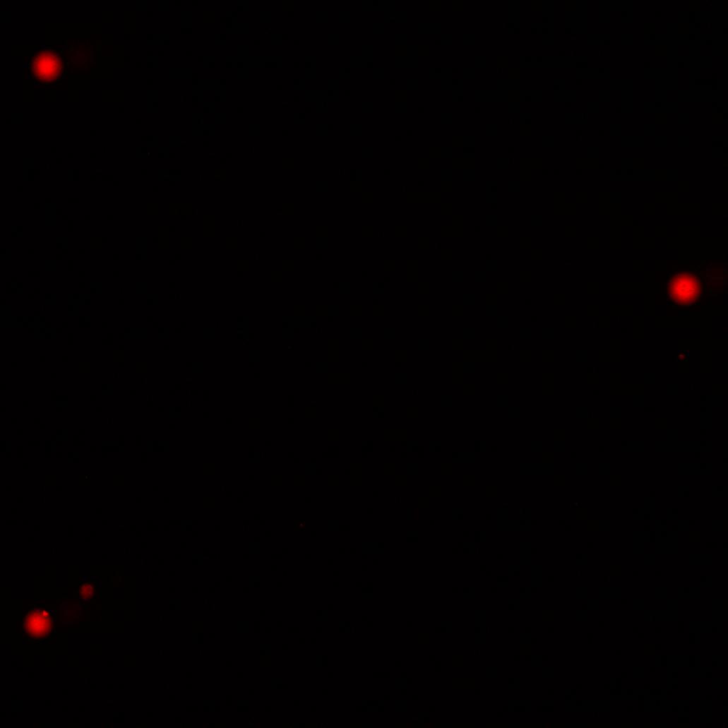
| + | When learning the basic techniques of synbio, we put together constructs for fluorescent protein expression in E. coli. We will return to GFP as a way to measure the solubility of reflectin, giving similar images to those below. | ||
| + | <gallery> | ||
| + | File:Cam JMFexperiment greencolonies.jpg | GFP in E.coli colonies. | ||
| + | File:Cam_positivecontrolexperiment_redcolonies.jpg | RFP in E.coli colonies | ||
| + | </gallery> | ||
| + | |||
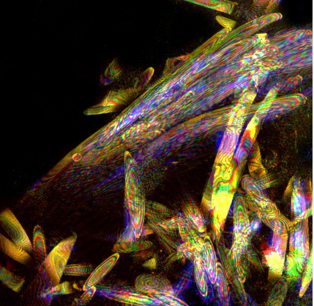
| + | We also experimented with unusual confocal microscopy settings to find a way of best capturing the iridescence and reflective properties of reflectin in cells. See below for images of squid cells we have studied. We hope to achieve similar images from E.coli cells. | ||
| + | |||
| + | <gallery> | ||
| + | File:Iridescent cells from squid eye.jpg | ||
| + | File: | ||
==In vitro photonic devices== | ==In vitro photonic devices== | ||
| Line 12: | Line 24: | ||
As part of our collaboration with nanophotonics, we have a number of techniques we plan to use in order to create different optical structures. For example, we aim make diffraction gratings, which will reflect light from across the spectrum (rather than the narrow range reflected by thin films). Reflectin appears to spontaneously form diffraction gratings on exposure to different solvents. We will also try layering reflectin with another polymer, to create a stacked structure which will give more intense colour due to additive effect of interference patterns. | As part of our collaboration with nanophotonics, we have a number of techniques we plan to use in order to create different optical structures. For example, we aim make diffraction gratings, which will reflect light from across the spectrum (rather than the narrow range reflected by thin films). Reflectin appears to spontaneously form diffraction gratings on exposure to different solvents. We will also try layering reflectin with another polymer, to create a stacked structure which will give more intense colour due to additive effect of interference patterns. | ||
| - | To build upon the work that has been done by researchers investigating reflectin, we hope to prove the feasibility of using these proteins as the next generation of flexible optical devices. Reflectin in cephalopods can give a range of colours based on dynamically changing the spacing of the protein assemblies, due to the charge differences caused by addition of phosphates. We hope to build a prototype of a non-backlit screen which reflects incident light of different colours. | + | To build upon the work that has been done by researchers investigating reflectin, we hope to prove the feasibility of using these proteins as the next generation of flexible optical devices. Reflectin in cephalopods can give a range of colours based on dynamically changing the spacing of the protein assemblies, due to the charge differences caused by addition of phosphates. We already know that the colour of a thin film can be altered by exposure to different solvent vapours (from blue to red, for example), and we are also planning to explore the interactions of reflectins with electricity in order to replicate the in vivo dynamism. |
| + | |||
| + | We hope to build a prototype of a non-backlit screen which reflects incident light of different colours, based on these potential methods for changing structural colour. | ||
Revision as of 13:33, 16 August 2011
Loading...
As this project is based around colour and light interactions, our characterisation experiments are set up to provide visual results in every facet of the project.
In vivo imaging
When learning the basic techniques of synbio, we put together constructs for fluorescent protein expression in E. coli. We will return to GFP as a way to measure the solubility of reflectin, giving similar images to those below.
We also experimented with unusual confocal microscopy settings to find a way of best capturing the iridescence and reflective properties of reflectin in cells. See below for images of squid cells we have studied. We hope to achieve similar images from E.coli cells.
==In vitro photonic devices==
|
We hope to build a prototype of a non-backlit screen which reflects incident light of different colours, based on these potential methods for changing structural colour.
|
==Previous iGEM projects==
|
|
As our project is a natural extension of previous Cambridge teams' efforts with light and pigment-based colour, we would be happy to show some of their finished projects alongside our own work.
|
 "
"


