 "
"
Team:Glasgow/BiofilmResults
From 2011.igem.org


Biofilm formation was also confirmed by SEM pictures that showed the extracellular matrix, such as Picture 1 below.
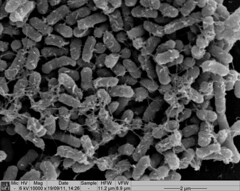 Picture 1: SEM image of Nissle biofilm showing the extracellular matrix |
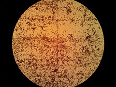
Picture 2: 400x A gram-stained 16-hour E.coli Nissle biofilm |
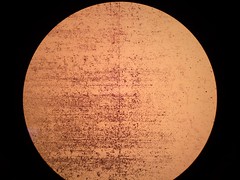
Picture 3: 100x A gram stained 16-hour E.coli Nissle biofilm |
The images below may show why E.coli Nissle has the biofilm forming that E.coli lab strains lack. In Picture 4 the fimbriae of Nissle are clearly visible. Since the E.coli lab strain in Picture 5 does not have them, they may be the reason E.coli Nissle 1917 are so adept at clinging to each other.
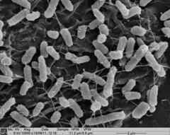 Picture 4: 10,000x SEM image of Nissle showing the fimbriae |
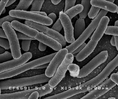
Image 2: 15,000x EM of E.coli for comparison. No fimbriae or EPS is visible. (courtesy of Rocky Mountain Laboratories) |