 "
"
Team:Glasgow/Biofilm/P. aeruginosa
From 2011.igem.org
| Line 44: | Line 44: | ||
<b>Figure 2: <i>Pseudomonas aeruginosa</i> biofilm growth over time.</b> These photographs were taken after 1hr, 14hrs and 48hrs of biofilm growth. They were stained using a Grams stain method that is designed specifically to avoid sheer forces being applied to the delicate biofilm structure. Details of this method are included in the <a href="https://2011.igem.org/Team:Glasgow/Lab_Book"><i>Pseudomonas aeruginosa</i> biofilms</a> lab book. | <b>Figure 2: <i>Pseudomonas aeruginosa</i> biofilm growth over time.</b> These photographs were taken after 1hr, 14hrs and 48hrs of biofilm growth. They were stained using a Grams stain method that is designed specifically to avoid sheer forces being applied to the delicate biofilm structure. Details of this method are included in the <a href="https://2011.igem.org/Team:Glasgow/Lab_Book"><i>Pseudomonas aeruginosa</i> biofilms</a> lab book. | ||
</br> | </br> | ||
| - | Go back to<a href="https://2011.igem.org/Team:Glasgow/Biofilm"> Biofilms</a> | + | <b><p>Go back to<a href="https://2011.igem.org/Team:Glasgow/Biofilm"> Biofilms</a></b></p> |
<h4>References</h4> | <h4>References</h4> | ||
<p>Flemming, H. & Wingende J., 2010. The Biofilm Matrix. Nature Reviews Microbiology 8, 623-633 </p> | <p>Flemming, H. & Wingende J., 2010. The Biofilm Matrix. Nature Reviews Microbiology 8, 623-633 </p> | ||
Revision as of 22:35, 21 September 2011


Pseudomonas aeruginosa
Pseudomonas aeruginosa is the standard organism for the investigation of biofilms, as it is very adept at forming them. This ability is mainly due to the production of exopolysaccharides (EPS), most notably the protein alginate, that is produced by the mucoid strains of P. aeruginosa and leads to the production of notably thicker and more stable biofilms,though it is not essential for biofilm formation
The strain that was chosen for this project is the wild type PA01 strain, that forms stable smooth, and more homogenous biofilms.
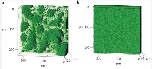
Biofilm architecture in muciod and non-mucoid P.aeruginosa strains. a) Typical biofilm architecture of the mucoid (alginate over-expressing) SG81 strain b)The smoother biofilm architecture typical of non-mucoid strains such as the wild type PA01 that was used in this project
It has long been postulated that P.aeruginosa has significant resistance to a wide range of antibiotics because of it's ability to form sturdy, resistant biofilms. Drenkard & Ausube showed that biofilm formation is strongly correlated to antibiotic resistance, allowing P.aeruginosa to thrive in almost any environment.
Properties of P. Aeruginosa Biofilms
|
Method
As we were trying to specifically disperse areas of biofilm it was necessary to establish a base rate of dispersal of the biofilm without using any of the dispersal mechanisms we designed. This would allow us to show quantitatively the increase in rate of dispersal that our different dispersal biobrick could generate when compared to a biofilm of non-transformed bacteria. To measure the base rate of dispersal glass slides were put into 50ml tubes containing 20ml of LB broth. The LB covered around a third of the glass slide, this is the area where the biofilm would form. The LB was then inoculated with 20μl of overnight culture of Pseudomonas aeruginosa. These tubes were then left on a bench top shaker at room temperature for a set amount of time (time points ranged from 1hr to 48hrs). After the biofilm had grown its allotted time the glass slide was carefully removed and placed into a fresh 50ml tube with 25ml of LB (which completely covered the biofilm) and left to allow the bacteria to disperse for 1hr. The slide was then transferred to a fresh 50ml tube with 25ml of LB. The biofilm is scraped off the slide using a thin flexible spatula. At this point both the dispersed cell and the biofilm scrapings were sonicated to stop clumping and plated in serial dilutions. |


Figure 1: Number of viable cells in biofilm compared to number of cells dispersed from the biofilm in one hour. The base rate of dispersal remains roughly proportional to the number of cells in the biofilm. The information for the time point hour 14 for the biofilm was not available. |
To make the images of biofilm formation, biofilms were formed on glass slides inserted into 50ml tubes. The tube was filled with 20ml of LB broth and inoculated with 20μl of over night culture of Psuedomonas Aeruginosa. The biofilms were left to form for the time indicated on the images.
Results The number of cells that dispersed from the biofilm seemed to be proportional to the number of cells in the biofilm with a ratio of roughly 5 dispersed:1 in biofilm. Figure 1 shows the number of dispersed cells when compared to the number of cells in the biofilm in both a graph and a table. Figure 2: Pseudomonas aeruginosa biofilm growth over time. These photographs were taken after 1hr, 14hrs and 48hrs of biofilm growth. They were stained using a Grams stain method that is designed specifically to avoid sheer forces being applied to the delicate biofilm structure. Details of this method are included in the Pseudomonas aeruginosa biofilms lab book.
Go back to Biofilms
References
Flemming, H. & Wingende J., 2010. The Biofilm Matrix. Nature Reviews Microbiology 8, 623-633
Drenkard,E. & Frederick M. Ausube